Enough (N)SAID about Ibuprofen & Soreness

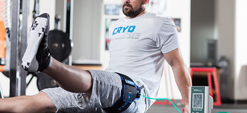
If I’m being honest here, it’s been a while since I’ve had a solid gym routine. But this semester I’ve been going pretty regularly, and let me tell you, I’ve felt the burn. My muscles have felt pretty sore in the 2-3 days following my workouts, so I’ve had to turn to ibuprofen a few times to relieve the pain. But even after taking ibuprofen in the morning, I’ve felt sore again by the end of the day. This got me thinking: how effective is ibuprofen at reducing muscle soreness?

Non-steroidal anti-inflammatory drugs (NSAIDs) are commonly served over-the-counter at pharmacies. Some common forms you may recognize include aspirin and ibuprofen (Motrin, Advil). NSAIDs are taken for many reasons; they reduce pain and inflammation, lower fevers, and reduce clotting action.[1,2] The typical dosage for adults who are looking to reduce mild-moderate pain is 400 mg every 4-6 hours. For adults who have pain caused by osteoarthritis, the typical prescribed dose is 1200 mg.[3] However, despite their pain reducing use, NSAIDs could yield negative side effects such as increased risk in developing nausea, stomach pains, or an ulcer.[1]

The mechanism of NSAIDs when it comes to reducing pain and inflammation is known and understood. After intense workouts, prostaglandins are produced by muscle cells. They aid in the healing process of muscle, but this often leads to inflammation, pain, and fever. Enzymes called cyclooxygenases (COX-1, COX-2) produce the prostaglandins that promote inflammation, pain, and fever. The goal of NSAIDs is to inhibit COX-1 and COX-2 from producing prostaglandins, thus decreasing the pain. However, the COX-1 enzyme is responsible for creating prostaglandins that protect the stomach lining and support platelet aggregation, so the inhibition of the enzyme is what could lead to stomach ulcers and the promotion of bleeding.[1,2,4] The science behind NSAIDs seems promising, but clinical research may prove otherwise.

Athletes commonly take NSAIDs after performing physical activity because they claim the drugs reduce pain and decrease recovery time. But here is the issue: only very few studies have been able to support this claim. Some studies have reported results that do indicate a beneficial effect, by stating NSAIDs used prophylactically mitigate exercise-induced inflammation, circulating creatine kinase levels, and muscle soreness.[5] On the other hand, these claims made by athletes lack scientific support. NSAIDs are known to treat inflammation, but many histological studies have proven that most overuse injuries are caused by tissue degeneration and not inflammation. Also, NSAIDs temporarily “mask” the pain caused by tissue degeneration or soreness. This does not ensure that muscles or tissues are actively getting healthier; it only hides the pain from the athlete. [5] Clearly, there are many different opinions about the use of NSAIDs, specifically ibuprofen, in the sports medicine field. Let’s take a look at what the “research says” about it. 

A study at the University of Saskatchewan was conducted to determine the effects of ibuprofen on muscle hypertrophy, strength, and soreness during resistance training. Participants (12 males, 6 females) trained their left and right biceps for six weeks, alternating arms on each day. The training program called for concentric curls at 70% of RM and eccentric curls at 100% of 1 RM. Every day after their training, they either received a 400 mg dose of ibuprofen or a placebo. On training days, each participant was asked to rate their soreness on a scale from 0-9. For both the placebo and ibuprofen, the participants reported soreness during the first week and that soreness decreased throughout the program to the point where participants felt no soreness in either arm during the final week. The researchers concluded that ibuprofen was not effective in reducing perceived soreness during the training. However, the researchers do not reflect on the limitations of their own study.  They had a small and uneven sample size when it came to gender and there could have been discrepancies and residual effects that came along with taking ibuprofen inconsistently. Additionally, they seemed pretty convinced by their findings, but maybe the dose they chose was not strong enough to show any reduction in soreness in a long term study.[6]

On the other hand, another study drew opposite conclusions. Researchers in Greece conducted a study to determine the effects of ibuprofen on delayed onset muscle soreness (DOMS) and muscular performance. Participants (14 men, 5 women) who have not done strength training in the last 6 months performed eccentric leg curls at 100% RM. Nine (9) subjects were given a 400 mg dose of ibuprofen every 8 hours for 48 hours after exercise, while the remaining 10 subjects received a placebo. The subjects rated their amount of soreness on a scale of 1-10 prior to exercising, 24 hours after exercising and 48 hours after exercising.  The results showed that muscle soreness was significantly lower for the ibuprofen group at both 24 hours and 48 hours after exercising. Similar to the previous study, the researchers did not evaluate the limitations of their study. The number of participants and number of each gender were low and uneven, respectively. Also, the soreness results were not discussed much in the conclusion of the paper. The researchers did not support why the soreness decreased with scientific evidence, which is what they did for the other the parameters they were testing for.[7]

Clearly, both studies came to different conclusions. However, both studies were conducted for different amounts of time, contained different exercises, and with subjects of different athletic abilities. There have been plenty of studies conducted to determine how effective ibuprofen is at reducing soreness, but each study contradicts the next. 

Overall, many studies show that ibuprofen is a short term solution to hiding muscle soreness, but it may not be effective long term. Though, I’m still going to keep on using it to treat my soreness.

Questions to consider:

  • Do you take NSAIDs to reduce your soreness after working out? How effective do you find them to be?
  • Do you think there’s a better way to measure soreness and how ibuprofen affects our muscles?
  • Do you think the length of the study has any correlation with the effectiveness of ibuprofen?

Sources: 

  1. (n.d.) Nonsteroidal Anti-inflammatory Drugs (NSAIDs). Retrieved from  https://www.medicinenet.com/nonsteroidal_antiinflammatory_drugs/article.htm#what_are_nsaids_and_how_do_they_work
  2. Tscholl, M., et al (2016). A sensible approach to the use of NSAIDs in sports medicine . Swiss Sports & Exercise Medicine , 65(2), 15–20.
  3. (n.d.) Ibuprofen (Oral Route). Retrieved from https://www.mayoclinic.org/drugs-supplements/ibuprofen-oral-route/proper-use/drg-20070602 
  4. (n.d.) What Are NSAIDs? Retrieve from https://orthoinfo.aaos.org/en/treatment/what-are-nsaids/
  5. Stuart J. Warden (2010) Prophylactic Use of NSAIDs by Athletes: A Risk/Benefit Assessment, The Physician and Sportsmedicine, 38:1, 132-138, DOI: 10.3810/ psm.2010.04.1770
  6. Krentz , J. (2008). The effects of ibuprofen on muscle hypertrophy, strength, and soreness during resistance training. Applied Physiology Nutrition and Metabolism , 33(3), 470–475. doi: 10.1139/H08-019

The Cold Facts on Icing

If you’re an athlete, there is a good chance that you have been told to ice your muscles after exercising. Icing is commonly thought to alleviate inflammation and soreness, as well as help to heal injuries caused by muscle overuse more quickly.1 There are different types of icing techniques popular in the world of athletics, ranging from a simple ice pack or frozen gel to cryotherapy and cold therapy chambers.2 Despite its wide use, there is some controversy regarding whether cold therapies are beneficial to the muscles or causing more harm than good.

Inflammation is an acute physiological response that is needed for tissues in the body to heal after exercising. Those opposing ice therapies claim that icing a sore muscle reduces its blood flow and slows the natural process of alleviating inflammation. While there is evidence that icing can help to reduce soreness in the short term after a workout, this reduction in the immune response can prevent the muscles from healing as quickly as they otherwise would.3 Many researchers have studied these countering views on the subject.


Figure 1. Cryotherapy machine [6]


One study aimed to see if topical cooling could improve recovery in eccentric contraction-induced muscle damage.4 They used a sample of 11 college male baseball players and put them into two groups; a control group and a group receiving topical cooling. The subjects used a barbell to complete 6 sets of 5 eccentric arm contractions. Those individuals in the cooling group received the ice 0, 3, 24,48, and 72 hours after the exercise for 15 minutes each. This was then repeated four weeks later. The researchers then analyzed the muscle hemodynamic changes, muscle damage markers, inflammatory cytokines, subject pain levels, and isometric muscle strength. The results showed that the subjects pain was similar between the two groups in the short term, but was greater in the later periods after the workout. The measured creatin kinase and myoglobin were significantly greater in the cooling group in the 48 and 72 hour periods than the control group. The cooling also resulted in higher hemoglobin concentration.4

Another study was conducted using 42 moderately active college aged males.5 The researchers had the subjects do 5 sets of 20 drop jumps, followed by lower body immersion in cold water. Three groups were used; one having a water temperature of 5 degrees Celsius, one with 15 degrees Celsius, and one control group. Measurements were taken on isometric knee extensor torque, countermovement jump, muscle soreness, and creatin kinase directly following exercise and 24, 48, 72, 96, and 168 hours after. The results for the countermovement jump showed that the warmer water group recovered more quickly than the colder water group. Creatin kinase remained elevated in all group except the warmer group, which returned to baseline at 72 hours. The subjects reported lower muscle soreness in the warmer water group as well.5

The research shows that icing sore muscles can be beneficial shortly after working out, but that people will possibly experience the same soreness later in time compared to people who don’t ice. It also makes it seem like using only slightly cold ice packs and water is more effective than using extreme cold. Athletes who ice should consider the amount of time they ice and the temperature they use when choosing cold therapies after a workout to avoid possible long term soreness and to improve with training.


Questions to Consider:

  • Do you think that using experienced athletes or people who only exercise occasionally was a more effective method of research?
  • Have your experiences with ice therapies been positive or negative?
  • What could a future study do differently to see the effects of icing on exercise?


References

  1. Cluett, J. (2019, September 25). How to Properly Ice an Injury. Retrieved from https://www.verywellhealth.com/how-to-ice-an-injury-2548842


  1. Gotter, A. (2017, February 2). Treating Pain with Heat and Cold. Retrieved from https://www.healthline.com/health/chronic-pain/treating-pain-with-heat-and-cold


  1. Aschwanden, C. (2019, February 5). Athletes love icing sore muscles, but that cold therapy might make things worse. Retrieved from https://www.washingtonpost.com/national/health-science/athletes-love-icing-sore-muscles-but-that-cold-therapy-might-make-things-worse/2019/01/31/a465dd84-1f25-11e9-8e21-59a09ff1e2a1_story.html


  1. Tseng, C.-Y. (2013). Topical Cooling (Icing) Delays Recovery From Eccentric Exercise–Induced Muscle Damage. Journal of Strength and Conditioning Research27(5), 1354–1361.


  1. Vieira, A. (2016). The Effect of Water Temperature during Cold-Water Immersion on Recovery from Exercise-Induced Muscle Damage. International Journal of Sports Medicine37(12), 937–943.


  1. (n.d.). 5 Cryotherapy Side Effects Therapists Should Watch For. Retrieved from https://www.homeceuconnection.com/blog/cryotherapy-side-effects-therapists/


Elevation Masks for Endurance Training: Stamina or Scam?

Endurance athletes across the globe are always looking for a way to gain an edge on their opponents. Some methods that have been adopted by elite and amateur athletes alike are altitude and respiratory muscle training. Altitude training involves training at high altitudes where oxygen is more limited than at sea level. Respiratory muscle training involves strengthening the muscles that are required for breathing. Both types of training involve creating a hypoxic condition for the body, meaning that the tissues are not receiving an adequate supply of oxygen. Exposure to hypoxic conditions stimulates the production of erythropoietin in the kidneys, which increases production of red blood cells. This creates an increase in the oxygen carrying capacity of the blood and has been correlated to an increase in endurance performance [1].These training techniques are said to increase aerobic capacity (VO2max), endurance, lung function, and overall performance in athletes [2]. 

Respiratory muscle training can be done using an elevation mask, which is designed to simulate the conditions of training at altitude while training at sea level (figure 1). Elevation masks cover the nose and mouth, restricting air flow and making respiration more difficult for the athlete. They often have values that allow for adjustments to the amount of oxygen that enters the mask. The Elevation Mask 2.0 by Training Mask LLC is one type of mask that uses values and can simulate altitudes ranging from 914 m to 5486 m [2]. But the question is – do these masks really cause physiological changes in the body to improve stamina and endurance?


Figure 1. The Elevation Mask 2.0 (Training Mask LLC, Cadillac Michigan) that can be used by athletes during training in hopes of improving performance [2]. It consists of a silicone mask and neoprene head strap, with adjustable resistance caps to change the amount of air flow.


Many studies have attempted to test these masks and determine if respiratory muscle training is actually beneficial to endurance athletes. Acclimating to high altitude occurs as the body increases the amount of red blood cells, which has been shown to improve sea-level running performance [1]. However, this hematological effect has not been consistently shown in studies that used elevation training masks. In addition to the volume of red blood cells, significant changes have not been observed in blood lactate concentration in people wearing the mask during training. These trends indicate that elevation masks may work as respiratory muscle training devices but do not accurately simulate the physiological changes that occur in the body at high altitudes [2]. 

Increased aerobic capacity, or ability to pump oxygenated blood to the muscles during exercise, is one of the main goals of endurance training. By participating in any sort of endurance training program, VO2max can be improved as the body adapts to the demands being placed on it. However, the goal of altitude and respiratory muscle training is to further enhance this ability to reach peak performance levels. Studies have shown that increases in VO2max for groups wearing a mask compared to increases in control groups are not significant [2]. In contrast, ventilatory threshold, which refers to the point during exercise where the rate of ventilation increases faster than the rate of oxygen uptake, and power output show a significant increase in experimental groups wearing a mask compared to control groups [3]. These findings indicate that wearing the elevation mask may help improve the function of the cardiovascular system during exercise.

Ventilatory threshold (VT) has been shown to correlate to the amount of work the muscles can maintain without fatigue. When the VT is surpassed, the muscles do not receive the necessary amount of oxygen and fatigue begins to set in. Therefore, increasing the VT for an endurance athlete should result in better performance [4]. In addition to endurance based metrics, respiratory muscle training has been shown to improve deep breathing and increase ventilatory efficiency throughout exercise[3,5]. 

Although there seems to be trends present in studies involving elevation masks and endurance training, there are limitations to what can be concluded. Most of the studies evaluated had limited sample sizes and the duration and intensity of the exercise regimes varied between studies. However, the studies do seem to imply that elevation masks may be beneficial to endurance performance through respiratory muscle training. By making it more difficult for the athlete to inhale and exhale, the body does appear to undergo physiological changes to adapt to the lower levels of oxygen. This adaptation may result in increased VO2max, VT, and power output over time. It seems that using an elevation mask does not cause any of the hematological changes in the body that occur when a person actually reaches a higher altitude. So although endurance performance may increase as a result of using the mask, it does not directly mimic the conditions of elevation training.


Questions to Consider:

  • Prior to reading this article, had you heard of professional athletes using altitude training or elevation masks to improve their performance? And if so, what sport did these athletes participate in?
  • Do you think amateur athletes and non-athletes could benefit from using an elevation mask in daily life?
  • Have you ever experienced altitude sickness? If so, what symptoms did you have?


References:

  1. de Paula, P., Niebauer, J. (2012). Effects of high altitude training on exercise capacity: fact or myth. Sleep Breath 16, 233–239. https://doi.org/10.1007/s11325-010-0445-1
  2. Porcari, J. P., Probst, L., Forrester, K., Doberstein, S., Foster, C., Cress, M. L., & Schmidt, K. (2016). Effect of Wearing the Elevation Training Mask on Aerobic Capacity, Lung Function, and Hematological Variables. Journal of sports science & medicine, 15(2), 379–386.
  3. Kido, S., Nakajima, Y., Miyasaka, T., Maeda, Y., Tanaka, T., Yu, W., Maruoka, H., & Takayanagi, K. (2013). Effects of combined training with breathing resistance and sustained physical exertion to improve endurance capacity and respiratory muscle function in healthy young adults. Journal of physical therapy science 25(5), 605–610. https://doi.org/10.1589/jpts.25.605
  4. Graef, J.L., Smith, A.E., Kendall, K.L. et al. (2008). The relationships among endurance performance measures as estimated from VO2PEAK, ventilatory threshold, and electromyographic fatigue threshold: a relationship design. Dyn Med 7(15). https://doi.org/10.1186/1476-5918-7-15
  5. Granados, J., Gillum, T., Castillo, W., Christmas, K., Kuennen, M. (2016). “Functional” Respiratory Muscle Training During Endurance Exercise Causes Modest Hypoxemia but Overall is Well Tolerated. Journal of Strength & Conditioning Research 30(3), 755-762.

Achey Breaky Muscles

Athletic expertise is not required for one to feel the aching and lasting effects of delayed-onset muscle soreness (DOMS). The aftermath of these effects can often exist along a spectrum ranging from significant muscle tenderness to debilitating pain.¹ In addition, the duration of these effects can exist along a spectrum ranging from 24 to 72 hours.¹ A common misconception correlates a good workout with the subsequent symptoms of DOMS, however, that is just not the case. With that being said, DOMS will often occur in accord with performing new exercises or increasing the intensity and/or duration of a current exercise.¹


The lingering soreness and stiffness as a result of DOMS can often cause individuals to seek methods that will ultimately reduce these symptoms and accelerate their return to the gym. Unfortunately, many of the treatment options on the market have mixed outcomes due to the individualized nature of exercise. Some may obtain adequate results through methods, such as massage or ibuprofen, while others may not; whereas, some methods prove to be completely ineffective, such as cryotherapy and stretching.¹ Furthermore, let’s give a look at a newly proposed treatment for DOMS and whether its results are promising.


Blood-flow restriction (BFR) training is often used at physical therapy as a passive way to regain strength without adding heavy weight. The venous blood supply is terminated at the area of injury, which in turn reduces the amount of oxygen at the site, activates anaerobic metabolism, and encourages muscle hypertrophy.² This proves to be an effective technique for restrengthening the muscle of patients who strictly lift lighter loads.² But, where is the crossover between BFR and DOMS?

Figure 1. BFR machine.[8]

BFR is thought to provide an alternative training method that will achieve similar muscle gains to resistance training with heavy loads while also minimizing the effects of DOMS. It has been postulated that BFR training may attenuate DOMS by preventing calcium-mediated proteolysis³ and by recruiting fast-twitch motor units.⁴ However, it has also been hypothesized that BFR training may induce muscle damage through ischemia-reperfusion (lack of oxygen in the tissues) or through the reduction of neutrophils that aid in inflammatory response.⁵


A 2019 study wished to examine the effects of BFR training on DOMS by using 25 untrained females and submitting them to isokinetic forearm flexion training, where half performed a bicep curl with a BFR cuff at a load reflecting a 30% eccentric-peak torque and the other half at a load reflecting a 30% concentric-peak torque.⁶ The findings revealed neither eccentric or concentric movements with a BFR cuff resulted in DOMS after the 7 days of training.⁶ These results were inconsistent with other studies that showed an increase in DOMS with concentric exercises.⁶ Is this inconsistency due to the low number of participants, the lack of diversity, or the duration of the experiment being only 7 days?


In contrast, a 2017 study compared the effects of BFR training and resistance training on DOMS by using 17 male participants and assessing their elbow-flexor muscle strength for 7 days in one of four training techniques: heavy load, light load, intermittent high-pressure BFR, and continuous low-pressure BFR.⁷ From this, the researchers found DOMS was significantly greater in BFR training (both intermittent high-pressure and continuous low-pressure) than in resistance training (both in heavy and light load).⁷


Much like the other methods of muscle recovery, BFR training may not be the most effective way to minimize DOMS. Exercise can be highly variable and strongly impacted by genetics, which may be the reason a consistent and reliable technique for treating DOMS has yet to be discovered.


Watch this youtube video to learn more about DOMS.⁸

Questions to consider

  • Based on the study mentioned in this article, do you think their findings are an accurate representation of the effects of BFR training on DOMS?
  • How have your opinions regarding DOMS changed with this post?
  • Did the video help you understand DOMS in an easily digestible way?


References

  1. Cheung K, Hume P, Maxwell L. Delayed onset muscle soreness: treatment strategies and performance factors. Sports Med. 2003; 33(2):145-164. DOI: 10.2165/00007256-200333020-00005
  2. Blood-Flow Restriction Training. American Physical Therapy Association. Website.http://www.apta.org/PatientCare/BloodFlowRestrictionTraining/. Updated May 24, 2019. Accessed February 20, 2020.
  3. Sudo M, Ando S, Poole DC, Kano Y. Blood flow restriction prevents muscle damage but not protein synthesis signaling following eccentric contractions. Physiol Rep. 2015;3(7):e12449.
  4. Loenneke JP, Fahs CA, Wilson JM, Bemben MG. Blood flow restriction: the metabolite/volume threshold theory. Med Hypotheses. 2011;77(5):748-752.
  5. Curty VM, et al. Blood flow restriction attenuates eccentric exercise-induced muscle damage without perceptual and cardiovascular overload. Clin Physiol Funct Imaging. 2017;38(3):468-476.
  6. Hill EC, Housh TJ, Smith CM, Keller JL, Schmidt RJ, Johnson GO. Eccentric and concentric blood flow restriction resistance training on indices of delayed onset muscle soreness in untrained women. Eur J Appl Physiol. 2019;119(10):2363-2373.
  7. Brandner CR, Warmington SA. Delayed onset muscle soreness and perceived exertion after blood flow restriction exercise. Journal of strength and conditioning research. 2017;31(11):3101-3108. doi:10.1519/JSC.0000000000001779
  8. Should You Still Train Sore? Youtube. Website. https://www.youtube.com/watch?v=Ut_4C_5CNbg. Published November 18, 2018. Accessed February 20, 2020.

Personalized Blood Flow Restriction. Owens Recovery Science. ORS. 2020c.

Dry Needling: Is it Worth the Pain?

Arriving at a physical therapy appointment to have a needle stuck deep into the body’s muscles only to leave hobbling and sorer than before doesn’t seem like an effective method for rehabilitation. However, the post-treatment benefits have made dry needling one of the many techniques individuals are using to treat and prevent injury from exercise.

What is Dry Needling?

While wet needling uses hollow needles to inject corticosteroids into muscle [7], dry needling (DN) consists of inserting a fine needle, similar to those used in acupuncture, deep into the muscle without injections. The needle is then twisted and moved around the area without being fully removed from the skin. The needling itself can be uncomfortable, feeling like a pinch, cramp, or deep prick, and can result in local soreness post-treatment. Physical therapists seek to insert the needle into a myofascial trigger point (MTrP) to relieve myofascial pain syndrome (MPS), the most common muscle pain disorder seen in clinical practice [1]. In exercise science, MTrPs are defined as “hyperirritable local point(s) located in taut bands of skeletal muscle or fascia which when compressed causes local tenderness and referred pain” [10]. Potentially caused by muscle overuse [2], this pain is commonly described as having a knot in a muscle and creates localized tenderness, pain to deep touch, and restricted movement [1].

The video above shows a physical therapist performing the dry needling technique on various muscles. Created by Dynamic Physical Therapy, Covington, LA (2013).

Dry needling is used as a rehabilitation technique to decrease the pain MTrPs can cause. The “fast-in and fast-out needle technique” applies high pressure stimulation to the MTrP, often causing a twitch response. These twitch responses are the result of a spinal reflex generated by the activation of nociceptors and mechanoreceptors. These receptors respond to the painful mechanical irritation and stretch the needle causes within the muscle [1]. When this occurs, a single motor unit fires and a visible, isolated contraction – the “twitch” – can be seen. These twitch responses can occur local to the needle or within muscles on the opposite side of the body. This phenomenon has led researchers to believe that the pain associated with MTrPs is due to central nervous system (CNS) changes [1]. 

How is Dry Needling Portrayed in Healthcare?

Healthcare providers, such as MedStar National Rehabilitation Network and ChristianaCare, have been advocates for dry needling. They mention DN is “an effective physical therapy modality…in the treatment of orthopedic injuries” [5] and that it can even be used for preventing pain and injury [4]. There have been many personal accounts of the wonders of dry needling in recovery from nagging injuries. AshleyJane Kneeland, who struggles with muscular pain due to lupus, fibromyalgia, and postural orthostatic tachycardia syndrome, cites DN treatment as relief for her painful spasms and headaches, as well as providing general relaxation [6]. But how effective is dry needling, really? Is there science to back up these claims?

What Does the Science Say?

Elizabeth A. Tough and co-authors performed a meta-analysis in 2009 of seven studies assessing the effectiveness of DN in managing MTrP pain. This study provides an update for the systematic review by Cummings and White, which found no evidence suggesting injections through wet needling generate a better response than dry needling [3]. One study found by Tough et al. suggests DN is more effective in treating MTrP pain than undergoing no treatment, two studies produced contradictory results when comparing DN in MTrPs to DN elsewhere, and four studies showed DN is more effective than other non-penetrating forms of treatment (placebo controls). However, when combining these studies for a sample size of n=134, no statistical significance was found between DN and placebo treatments. 

While the authors conclude the overall direction of past studies trend towards showing that DN is effective in treating MTrP and MPS [10], there is no significant evidence yet. The lack of statistical significance could be due to low consistency in study design for studies included in the meta-analysis, as each employed varying mechanisms for needle placement, depth, and treatment frequencies, along with there being an overall small sample size. Therefore, further studies are required to significantly conclude that DN is effective in MTrP rehabilitation.

Ortega-Cebrian et al. recognized the limitations in previous studies and thus sought to create a significant evaluation of the ability of DN to decrease pain and improve functional movements. The authors use a myometer (MyotonPro, [8]) and surface electromyography (sEMG) to assess the mechanical properties of muscle in subjects (n=20 M) with quadricep muscle tension and pain [9]. 

The MyotonPro allows researchers to quantify muscle tone and stiffness. While no standards exist for describing these parameters with respect to changes after rehabilitation techniques, researchers found the device to be reliable through inter-rater reliability (comparing values of the MyotonPro to another rater). Pain was assessed by subjects using the Visual Analogue Scale (VAS) and a goniometer was used to measure small range of motion (ROM) improvements. DN was performed by one of two experienced therapists until twitch responses ceased [9].

Authors report that DN resulted in statistically significant pain reduction and an increase in flexion ROM. However, the ROM was very small and could be within the range of measurement error of the goniometer. Also, the p-values reported in-text for these parameters do not match the corresponding table which presents a question of the reliability of author reporting. All sEMG parameters, except for decreased vastus lateralis activity, were not significantly changed by DN, as well as all MyotonPro parameters, besides a decrease in vastus medialis decrement (muscle elasticity) and resistance. In a power analysis performed after the study, authors report needing 198 subjects for statistically significant results – much higher than the 20 subjects used [9]. Therefore this study continues the uncertainty in the benefits of DN, but does present significant subject-reported pain reduction.

Is it Worth the Pain?

So is dry needling worth the pain? After being put to the test through experimental studies, there is no clear evidence that dry needling is more beneficial than alternative rehabilitation methods such as wet needling, placebo needling, or acupuncture [9]. However, while the mechanisms of changes in muscles with trigger points due to dry needling are unknown, subjects do report pain reduction. Dry needling should be taken on a case-by-case basis since current knowledge of widespread benefits is limited. Essentially, if dry needling treatment alleviates pain more than other rehabilitation methods and the pain of the procedure is bearable, why not give it a try?


Questions to Consider:

  • Would you be willing to try dry needling regardless of uncertainties in the literature?
  • Do you believe it is a problem that healthcare providers claim dry needling is effective despite a lack of conclusive evidence?
  • What should future studies do to ensure significant results?


References:

[1] Audette, J. F., Wang, F., & Smith, H. (2004). Bilateral Activation of Motor Unit Potentials with Unilateral Needle Stimulation of Active Myofascial Trigger Points. American Journal of Physical Medicine & Rehabilitation, 83(5), 368–374. doi: 10.1097/01.phm.0000118037.61143.7c. 

[2] Bron, C., & Dommerholt, J. D. (2012). Etiology of Myofascial Trigger Points. Current Pain and Headache Reports, 16(5), 439–444. doi: 10.1007/s11916-012-0289-4. 

[3] Cummings, T., & White, A. R. (2001). Needling therapies in the management of myofascial trigger point pain: A systematic review. Archives of Physical Medicine and Rehabilitation, 82(7), 986–992. doi: 10.1053/apmr.2001.24023. 

[4] Dry Needling®. (n.d.). Retrieved from https://christianacare.org/services/rehabilitation/physicaltherapy/dryneedling/

[5] Dry Needling. (n.d.). Retrieved from https://www.medstarnrh.org/our-services/specialty-services/services/dry-needling/

 [6] Dry Needling: The Most Painful Thing I’ve Ever Loved. (2015, March 25). Retrieved from https://www.everydayhealth.com/columns/my-health-story/dry-needling-most-painful-thing-ever-loved/

[7] Dunning, J., Butts, R., Mourad, F., Young, I., Flannagan, S., & Perreault, T. (2014). Dry needling: a literature review with implications for clinical practice guidelines. Physical Therapy Reviews, 19(4), 252–265. doi: 10.1179/108331913×13844245102034. 

[8] Muscle Tone, Stiffness, Elasticity measurement device. (n.d.). Retrieved from 

 [9] Ortega-Cebrian, S., Luchini, N., & Whiteley, R. (2016). Dry needling: Effects on activation and passive mechanical properties of the quadriceps, pain and range during late stage rehabilitation of ACL reconstructed patients. Physical Therapy in Sport, 21, 57–62. doi: 10.1016/j.ptsp.2016.02.001. 

[10] Tough, E. A., White, A. R., Cummings, T. M., Richards, S. H., & Campbell, J. L. (2009). Acupuncture and dry needling in the management of myofascial trigger point pain: A systematic review and meta-analysis of randomised controlled trials. European Journal of Pain, 13(1), 3–10. doi: 10.1016/j.ejpain.2008.02.006.

Kinesio Tape : Does it Really Work?

Most individuals, athletes or not, have experienced a musculoskeletal injury due to the overuse of a specific tissue or muscle. These overuse injuries can slow down an individual either in the workout routines or daily life. While not all injuries react the same way, many overuse injury areas are known to build up lymphatic fluid causing swelling and pain. The swelling and pain come from the accumulated lymphatic fluid putting increased pressure on the injured muscle or tissue.


Taping using Kinesio Tape (KT) has become a very popular proposed treatment and recovery aid over the past couple of years. KT became popular after the 2008 Beijing Olympic games, where beach volleyball player Kerri Walsh Jennings caught the attention of many spectators for wearing multi colored tape strips on her shoulder. KT is believed to lift the skin from the underlying layers of fascia, or bands or connective tissue. The lifting of the skin from the fascia results in a greater movement of lymphatic fluid, which transports white blood cells throughout the body and removes bacteria, waste products, and cellular debris. When the tape is correctly used it may also be able to provide support to the surrounding muscles and help to ensure that the muscle does not over extend or over contract [1].


Figure 1. Athlete wearing Kinesio Tape.


Research suggests show that the tape will allow increased oxygen to the injured muscle and decreased inflammation. A 2012 study tested the effects of KT on blood flow in the gastrocnemius muscle and whether or not the way KT is applied changes the outcome on the muscle performance. In this study 61 healthy active individuals with no recent leg injuries were assigned to either treatment KT, sham KT, or a control group. Before taping a blood flow, circumference, and water displacement was taken for the gastrocnemius muscle. The individuals were then taped, and each measurement was taken again 24 hours and 72 hours after being taped. The results of this study showed no significant differences in the blood flow to the muscle using KT. There was also no change in the muscle’s performance based on the application technique of the tape [1].


From five previous systematic reviews, a new systematic review had been created to evaluate whether or not KT was more effective than no treatment or a placebo treatment, for people with musculoskeletal conditions, on pain levels, disability, and quality of life. Several different studies had been performed that looked at the pain levels on a scale from (0-10) for performing different activities while wearing either KT or another form of tape. These studies are prone too potential bias from the users and small sample sizes. Many of the referenced studies only shared certain of the results or were considered significant but of low quality [2].


Within a study done on subjects who had been diagnosed with rotator cuff tendonitis/impingement similar results were found as in the studies before. The only difference in this study was that they took self-reported measurement for range of motion along with pain. While the taping was ineffective compared to sham tape in long term, the KT provide immediate in pain free abduction range of motion. Once again, this study was limited to a. young population and it lacked a control group for comparison [3].


Although studies show that KT is ineffective in aiding injury rehabilitation, it is. Still used often by many groups of people. Since KT is relatively safe there is no reason why it cannot be used. Whether or not KT acts as a placebo or works I ways that are yet to be understood, it has worked for a large population of people for many years in helping to get past injuries for exercise and daily life.


Questions to Consider


Have you ever used Kinesio Tape? If so, did it help alleviate pain or support movements?


KT placebo effect or valid injury rehabilitation aid?


Do you think KT will last as an injury aid?


References


[1] Hannah L. Stedge, Ryan M. Kroskie, and Carrie L. Docherty. (2012). Kinesio Taping and the Circulation and Endurance Ratio of the. Gastrocnemius Muscle. Journal of Athletic Training, 47(6), 635-642.


[2] Patricia do Carmo Silva Parreira, Luciola da Cunha Menezes Costa, etc. (2014). Current evidence does not support the use of Kinesio Taping in clinical practice: a systematic review. Journal of Physiotherapy, 60(1), 31-39.


[3] Mark D. Thelen, James A. Dauber,  Paul D. Stonemen. (2008).Journal of Orthopaedic & Sports Physical Therapy,38(7), 389-395.


[4] “WHAT’S THE DEAL WITH THE TAPE? Benefits of Kinesiology Theraputic (KT)Tape-Small Tool Delivers Big Impact.” Fischer Institute, 16 Oct. 2017, fischerinstitute.com/benefits-kinesiology-therapeutic-tape/.


Acute/short-term effects of stretching


Stretching is a critical component of many regimens seen in clinical and fitness settings. Whether you’re a person who prefers to stretch before/after your routine, many people will attest to the physiological benefits of stretching. Proponents of stretching believe that it improves performance during exercise and prevents injuries and soreness. Some would go so far as to say that an individual may not be stretching enough when they repeatedly experience pain or injury after their workouts with no signs of improvement. Despite these enduring beliefs, the science behind the benefits of stretching is questionable. For the purposes of this blog post, we will focus on the acute, short-term effects of stretching on performance during exercise.

Three forms of stretching  used in exercise and rehabilitation settings include dynamic stretching, ballistic stretching, and static stretching. Dynamic stretching is a type of stretching which involve fluid-exaggerated movements. Ballistic stretching utilizes fast countermovements. Static stretching involves extending target muscles to a limit point, and maintaining that position for an interval between 10 and 30 seconds. In order to minimize injuries, static stretching is encouraged for non-athletes.

Numerous scientific studies have shown that have shown that static stretching results in an improved joint range of motion  (ROM) and greater flexibility in the muscles targeted by this technique. Conversely, research has also shown that stretching before exercises can result in a lower force output generated in the muscles that are targeted. Compliance is the lengthening of muscle fibers in response to an applied force. According to an article cited by the the National Institute of Health (Anderson, 2005), increased compliance (which occurs a result of stretching) has been linked to a decreased ability to absorb force at rest, whereas decreased compliance results in a muscle being able to withstand higher tension. This is significant because, when sarcomeres are stretched to the point that the actin and myosin filaments do not overlap, the force absorbed is transmitted to the muscle fiber cytoskeleton; resulting in fiber damage (regardless of a muscle’s joint ROM). Thus, compliance may result in decreased performance depending on the type of exercise performed. Another issue that arises related to the use of stretching before exercise is the type of stretching utilized. Science has shown that muscle fibers can experience tension when stretched as little as 20% of their total length1. Thus, it is difficult to establish a universal standard describing correct stretching techniques. In addition, improved joint ROM can be attributable to extraneous factors (such as increased pain tolerance); making the strength of its relationship to stretching highly questionable.

There are a plethora of studies conducted that attempt to quantify the effect of stretching on performance. One study, conducted by researchers at Sahmyook University in 20182 examined the effects of stretching on muscle strength, endurance, and endurance in a non-athletic sample of 13 active collegiate male students. These subjects were separated into three groups: those who did not perform any warm ups before exercise  (NWU), those who performed aerobic warm ups in the form of power walking for ten minutes (AWU) before exercise, and those who performed aerobic warm ups with static stretching for ten minutes (ASU). All three groups performed isokinetic muscle testing. The stretching used in the study consisted of straddling, seated calf stretching, and standing quadriceps stretching for the lower body. Two repetitions of each stretching motion were performed for 20 sec each and the entire stretching program took 5 min to perform. All subjects rested for 1 min after warming up and then underwent isokinetic muscle testing of the knee joints. The sequence of performance of each warm-up exercise was individually randomized. In the successive weeks, each group was tested according to the type of warm-up performed. The testing was conducted for 3 weeks, and all groups were allowed a week to rest in between tests.

In order to quantify the results in each group, a knee extension/flexion isokinetic  dynamometer was used. Participants were asked to extend and flex the knee by exerting their maximum strength as fast as possible while keeping their trunk up against the backrest during the test and to hold onto the handles. The subjects performed the maximal test of four repetitions. Each maximal test was conducted with an angular speed of 60°/sec to measure isokinetic muscle strength and an angular speed of 180°/sec to measure isokinetic muscle power. In addition, the muscle endurance test was conducted with an angular speed of 240°/sec. The exercise was conducted twice prior to testing to familiarize the subjects with the test, thereby achieving optimal results. The subjects were verbally encouraged and allowed to view their torque graphs during testing as a form of visual feedback to increase motivation.  To analyze muscle strength, power and endurance, measurements of the left and right knee joints were divided into each independent variable before data processing was performed. In addition, psychological evaluations in the form of questionnaires were administered to subjects before and after workouts for individuals in all three groups. These assessments utilized a 5-point Likert scale (1, very bad; 2, bad; 3, average; 4, good; 5, very good). The Kruskal–Wallis rank test were used to examine the differences of variables among groups and the Wilcoxon test was used to investigate psychological conditions before and after warm-ups within times in each group. A Mann–Whitney post hoc test was implemented to detect any significant differences in the Kruskal–Wallis test. The significance of all data was established at p ≤0.05. The results from the table have been included in figures attached to this post. The data is shown in the bottom of this point via a hyperlink. 

Based on the results of this experiment, the researchers concluded that there was no significant effect of the type of warm-up activity on performance in any of the tests performed in this study. Shown in Table 2, at 60°/sec (which is an angular speed for rating muscle strength), the NWU showed higher rates for both the extensor and flexor. However, the researchers determined that the difference was not statistically significant Shown in Table 3, at 180°/sec (an angular speed associated with rating muscle power), AWU and ASW groups attained higher rates for the flexor and extensor, respectively, although the difference was not statistically significant. The total work at 240°/sec (which reflects muscle endurance) was higher in ASW for both the flexor and extensor than NWU and AWU, though not statistically significantly. These results are shown in Table 4. In a similar manner to the trends seen when evaluating athletic performance, the individuals in the ASW group marked higher scores on their psychological assessments than the AWU and NWU groups. The results are shown in Table 5. However, the researchers determined that the result were not statistically significant.

Overall, while there appears to be some merit to the psychological benefits of stretching before exercising, its effect on athletic performance remains inconclusive. However, if you find that stretching helps improve your outlook/state-of-mind during the course of your workout, I would highly encourage you to continue your routine.


Questions to Consider

  1. Based on the experiment, do you believe that stretching before a workout provides any benefits/advantages towards performance?
  2. Does this post affect your views towards stretching?
  3. Would you encourage someone seeking to exercise more frequently to stretch before/after their exercises?


References

  1. Andersen JC. Stretching before and after exercise: effect on muscle soreness and injury risk. J Athl Train. 2005;40(3):218–220.
  2. Park HK, Jung MK, Park E, et al. The effect of warm-ups with stretching on the isokinetic moments of collegiate men. J Exerc Rehabil. 2018;14(1):78–82. Published 2018 Feb 26. doi:10.12965/jer.1835210.605

Results

I wanna rock and roll (out my myofascial tissue)!

Anyone who has partaken in any physical activity, whether it is a sport, exercise routine, or just simple around the house chores that require a little more muscle power than normal, has experienced muscle soreness or discomfort.  Generally, when muscles are pushed passed what they are used to (i.e. new exercising routines, increased weight, eccentric exercises) the muscle fibers undergo damage and the body’s response is to add muscle fibers and/or to increase the size of muscle fibers to help increase muscle strength. When talking about sore muscles, it is generally thought the soreness comes from the physical effects of muscle tearing, repairing, and growing from a workout. There is, however, one part of the muscle that plays a role in not only soreness, but also range of motion (flexibility), and muscle performance that not many people know needs special attention: the myofascial tissue.

Figure 1: Skeletal muscle structure through different layers of the muscle. Myofascial tissue lies over the epimysium connective tissue which coats the muscle bundle. The epimysium, perimysium, and endomysium are specialized versions of myofascial tissue.

Different forms of fascia can be found all over the body, from encasing organs, to blood vessels and nerves, to muscle.  Fascial tissue that specifically covers muscle, or myofascial tissue, is a thin, white/transparent connective tissue that covers muscle, bundles, muscle fibers, and the muscle as a whole.  If you have ever picked off the thin white stuff covering parts of a chicken breast while preparing it for dinner, you tore off the myofascial tissue layer. Myofascial tissue is an extremely flexible and strong material, which is made up of elastin fibers, for stretch, and collagen fibers, for strength, that are embedded in a gelatinous ground substance, which reduces friction between the muscle fibers and promotes ease of motion [1]. Considered a “deep fascia,” myofascial tissue is made up of a more compacted weave than other fascia found throughout the body and can modify itself depending on the forces placed on it.   (Figure 2).  Because of this, if there is “trauma” or “injury” to the tissue, it can become out of alignment and

Figure 2: 3D visualization of myofascial tissue (white web like structure) and fascia tissue between the skin and muscle (yellow web like structure). Myofascial tissue can be related to a cotton candy structure that is extremely complex and strong. Retrieved from https://www.myofascialrelease.com/about/definition.aspx

cause trigger or dysfunctional points. These points, most commonly referred to as knots, is when the fibers that make up the tissue gets stuck together, loses its elasticity, and becomes taught [2]. Polly de Mille, R.N., C.S.C.S., director of performance services at the Hospital for Special Surgery in New York City explains in an interview for SELF that it is very similar to getting ice cream in silky smooth hair.  When there are “knots” in the muscle fascia, it limits range of motion, and can trigger immune responses which can ultimately lead to pain and discomfort (cytokines have been shown to cause pain and soreness) [3,4].


Now, what is the best way to heal and prevent muscles from experiencing these knots and discomfort? When talking about getting rid of knots in your body, a massage should be the first thing that comes to mind.  The “hurts so good” mentality of deep massaging muscles to where the patient feels pain and then relief afterwards is a popular desire, though not for everyone. Applying pressure and different forces to the tissue through a massage, or foam roller which we will talk about in a little bit, while moving around the fascia helps to separate and relax the tissue and muscle, allowing it to go back to its natural state.  Effects also include an increase in blood flow, which should help muscles get the proper nutrients to repair. Massaging also releases “feel good” brain chemicals, like endorphins, which basically inhibit pain receptors and overall makes you feel better. A study conducted by Mal-Soon Shin and Yun-Hee Sung induced muscle fatigue on 21 young males and treated 11 of them to massages afterwards while recording surface muscle activation and position of their medial gastrocnemius muscle.  According to their study, massaging increases muscle activation and strength due to a change of structural properties. However, in their discussion, they mention that not all messages are effective, which seems to be a common issue in the argument of whether foam rollers, or self myofascial release in general, works or not [5].

Massages are so great because when another person is working out your muscles, they are not only more accurate in pinpointing the location, but they can also apply more force (remember: collagen is EXTREMELY strong in ratio to its size. Proportionally, it is stronger than steel!).  As great as they are, unless someone at home is a masseuse, it can be costly. Self myofascial release techniques, such as foam rolling, have taken over the exercise world and are now regularly used. Foam rolling is when the user applies pressure to “trigger point” or sore spot before or after a workout by using their body weight to roll against a foam cylinder. Though it feels good, does it actually work?

Research has proven that foam rolling is great for warming up muscles and increasing range of motion and flexibility, but the verdict is still out on decreasing muscle soreness.  Though the mechanism behind foam rolling is not exactly known, there is great evidence that it does work on some type of level, whether it is physical or just simply mental. In a systematic literature review of research on using a foam roller before and after workouts, Scott W. Cheatham identified different scientific articles that were critically appraised with trusted conclusions.  From these articles, he identified five studies on the effects of foam rolling and range of motion before exercising. All of these studies resulted with an increase in stretching or range in motion in test subjects [6]. It is common knowledge to stretch before a workout or game to help “warm up” the muscles so that they’re are more flexible, which helps prevent injury and soreness. It is also speculated that rolling out could create a friction that literally heats up the fascia and muscle, making it more flexible and the typical “loose” feeling [2].  After a workout, however, there is a preconception that rolling out will help with delayed onset muscle soreness and pain in general. In this literature review, Cheatham identifies two different journals that conclude that foam rolling does reduce pain, but since the mechanism behind it is still unknown, how much can we trust? In the same SELF article as mentioned previously, Lewis J. Macgregor, Ph.D., an exercise physiologist and lead author of the University of Stirling confirms foam rolling does help increase blood flow, which in turn promotes muscle recovery, but foam rolling does not actually help with myofascial release. Since the collagen in the fascia is so strong, it is argued using your body weight to roll out is not enough. Instead, the pressure of rolling stimulates nerve receptors, which sends the same “hurts so good” feeling to your brain that is then perceived as loosening up the muscle, when really it is not happening.

Overall, foam rolling and myofascial release is an effective way to warm up your muscles and stretch them out before a workout and to help stimulate more blood flow post workout, just don’t get your hopes up about avoiding soreness! Increasing range of motion and flexibility before a workout is a great step to ease muscle soreness, but foam rolling alone is not the answer.  The verdict is still out on the mechanism behind the effects of myofascial release on a cellular level, but hey, if it feels good, why not!


If you’re interested, here is a video of foam rolling techniques because like anything, it’s not effective if you don’t do it properly.

https://www.youtube.com/watch?v=WCj1dvTwOF0


Questions to consider:

Is it worth it to foam roll or stretch in general before physical activity?

Is myofascial release something to think about daily and not just dealing with exercising?

Do you think foam rolling has a placebo effect or is it both systems (nervous and skeletomuscular) working together?

Do you think foam rolling is just another exercising fad?

Would adding heat be beneficial? Or a waste of time?


Sources:

  1. Shah, S., & Bhalara, A. (2012). Myofascial Release. International Journal of Health Sciences and Research,2(2), 69-77.
  2. Fetters, K. A. (2018, July 21). Here’s What Foam Rolling Is Actually Doing When It Hurts So Good. Retrieved April 12, 2019, from https://www.self.com/story/what-foam-rolling-is-actually-doing-when-it-hurts-so-good
  3. Grosman-Rimon, L., Parkinson, W., Upadhye, S., Clarke, H., Katz, J., Flannery, J., … Kumbhare, D. (2016). Circulating biomarkers in acute myofascial pain: A case-control study. Medicine, 95(37), e4650. doi:10.1097/MD.0000000000004650
  4. Zhang, J. M., & An, J. (2007). Cytokines, inflammation, and pain. International anesthesiology clinics, 45(2), 27–37. doi:10.1097/AIA.0b013e318034194e
  5. Shin, M., & Sung, Y. (2015). Effects of Massage on Muscular Strength and Proprioception After Exercise-Induced Muscle Damage. Journal of Strength and Conditioning Research,29(8), 2255-2260. doi:10.1519/jsc.0000000000000688
  6. Cheatham, S. W., Kolber, M. J., Cain, M., & Lee, M. (2015). THE EFFECTS OF SELF-MYOFASCIAL RELEASE USING A FOAM ROLL OR ROLLER MASSAGER ON JOINT RANGE OF MOTION, MUSCLE RECOVERY, AND PERFORMANCE: A SYSTEMATIC REVIEW. International journal of sports physical therapy, 10(6), 827–838.

Whole Body Air Displacement Plethysmographic- Problem

PROBLEM

In order for whole body air displacement plethysmographic machines such as the BodPod to function optimally (so that viable data can be collected), it is crucial that laminar flow is maintained throughout the machine’s ventilation system at all times. Imagine that you are an engineer (imagine that!)  tasked with manufacturing the tube components for the Bod Pod.

If the flow rates in the inlet and outlet tubes are equal, the volumetric  flow rate of air in the tubing system will be 0.25 cubic meters/second , and the BodPod functions in laminar/laminar-like conditions, what are the ideal dimensions for the diameters of the inlet and outlet tubes in the Bod Pod?


Assumptions

  • Flow rates are equal in the inlet and outlet tubes
  • The tubes are cylindrical  
  • Laminar flow is maintained at all times
  • Pressure changes are negligible
  • Air circulating inside the BodPod has similar thermodynamic/kinematic properties ambient air at room temperature
  • Temperature conditions of the device are identical to those at room-temperature

A link has been included to a power point presentation that contains diagrams that will aid readers in solving this problem:

https://docs.google.com/presentation/d/178RDbb09Vjwv3o7JVS9bkwR1TMjgWWApOBE5WDRzaQE/edit#slide=id.p


Figure 1: Schematic of Adult-Sized Bod Pod and circuitry components that will be used as a reference for this problem.


BACKGROUND KNOWLEDGE/ ASSUMPTIONS

According to the 4th page of the patent filed by the manufacturer, Life Instruments Inc., it is okay to assume laminar conditions inside the tubing ventilation due to the fact that flow rate inside the inlet and outlet tubes are always set to values of low magnitudes. Literature in courses such as Signals and Systems show that low flow rates result in low generation of acoustic noise by  air circulation systems.


I was unsuccessful in locating some sort of testing standard that establishes set values for the volumetric flow rates of air in laminar conditions. There appears to be any information pertaining to any testing protocols the manufacturer used for design verification purposes in the original 510(k) form filed with the FDA. To establish an appropriate flow rate value for this test question, I searched for similar problems online. In short, the values for the volumetric flow rate of air (Q) ranged from 0.1 to 0.8 cubic meters/second in my searches. I decided to use a value of 0.25 cubic meters/second in this problem. By assuming that the values for Q are equal for both tubes, it is possible to design both tubes with an equal diameter. Thus, along with other reasons that will be outlined later in this section, all the solver is required to do to calculate the correct value in this problem is to use one equation.

Normally, pressure fluctuations trigger changes in tubings and pipes create flow gradients in closed ventilation systems. Because of this, mathematical expressions such a Boyle’s Law and Bernoulli’s equations are used to solve changes in volume and volumetric flow when pressure fluctuations occur. According to page 4 of the patent filed for the Bod Pod, the authors state that the use of pressure transducers which are coupled to the inlet and outlet tubes helps monitor any pressure changes that occurs in the tubing; automatically adjusting the pressure settings in the tubes to more optimal levels through negative feedback. This is done in order to maintain a constant flow rate (and thus, laminar flow throughout the circulation system). Later on in section 4 of the patent,  the manufacturers also state that constant air flow can be maintained with the addition of rotary pumps to the circulation system (which are not actively displayed in any of the figures included).

The manufacturer’s statements in the patent confirm the presence of temperature-sensing circuitry in the inlet and outlet tubes that control the internal temperature of the environment inside the tubing and the pod itself. Thus, any temperature fluctuations that could create flow gradients in the device’s tubing are negligible since they are always corrected in  rapid fashion. This also eliminates the need for Fourier’s law to solve the value of Q in this problem.

Assuming that the tubing is cylindrical eliminates the need to solve for any hydrodynamic radius  values(which are used in equations associated with fluid flow in which tubes/pipes are any shape that is non-cylindrical).


By assuming that the air inside the device’s circulation system behaves in a similar fashion to ambient air, and that the conditions inside the circulation system are similar to those at room-temperature and that the device is used in STP conditions, it is possible to estimate the value of the kinematic viscosity of air (which is needed to solve the value for the diameter of the tubing using the Reynolds number equation along with the value of the flow rate given in the problem description and the upper-limit value of the Reynolds number associated with laminar flow).


SOLUTION


In order to solve for the value of the tube diameter, the solver must utilize the following equation:

Re = QD/v ,

Re = reynolds number

Q = volumetric flow rate of air

D = pipe bore or tube diameter

v = kinematic viscosity

Reynolds number flow rate equation-16umdck  <— Click the link to view a more detailed image of the equation


NOTE: Pipe bore is equivalent to the diameter of the tube, and this equation is applicable to both pipe and duct installations.

First, the value of Q is already provided in the description. So the reader is already provided one unknown.

Second, the reader is told in the problem description and background section to assume laminar conditions in the circulation system. The Reynolds number value used in this problem is 2300, which is the established upper limit for laminar flow. All values at or below this number is considered laminar flow.

Third, since the reader is told to assume that the air circulating through the inlet and outlet tubes are similar in kinematic/thermodynamic behavior to ambient air at room temperature, the reader can assume that air inside the circulation system has the same kinematic viscosity as ambient air at room temperature. This value is 1.494 x 10⁻⁵ meters ^2/ second.

At this point, the only unknown that the reader is left with is the value of D, or the tube diameter. After plugging all the known values into the above-aforementioned equation and solving for the value of D algebraically, the reader should arrive at a diameter value of approximately  0.13708 meters.


REFERENCES


[1] Dempster Phillip, Michael Homer, and Mark Lowe (2004). United States Patent 20040193074A1. Retrieved from https://patentimages.storage.googleapis.com/93/cf/ea/6d2d1346ea1129/US20040193074A1.pdf


[2] Engineers Edge. “Kinematic Viscosity Table Chart of Liquids” (2019). Machinery’s Handbook, 29th edition.  Retrieved from

https://www.engineersedge.com/fluid_flow/kinematic-viscosity-table.htm


[3] Foster, Trevon. “Laboratory Flow Meters: Flow Measurements In the Lab” (2015). Titan Enterprises, Ltd. Retrieved from


https://www.flowmeters.co.uk/laboratory-flow-meters-flow-measurement-in-the-lab/

Resistance Temperature Detector Calibration for Sweat Sensors

Glucose and Lactate are two analytes in sweat that would be highly desirable to apply sweat sensing technology to, each for their own individual reasons. Since the biosensing technology typically used to detect these analytes utilizes enzymatic reactions, temperature of the sample being tested must be taken into consideration when interpreting results due to its effects on enzymatic activity. Therefore, temperature sensors are an essential component of any sweat sensor that aims to give reliable feedback on either/both of these analytes. Multiple temperature sensing technologies exist, but a simple, commonly used technology is resistance temperature detectors (RTDs). These simple circuits use a Wheatstone bridge with a pure metal resistor that is exposed to the sample being tested. That resistor has a temperature-dependent resistance, and its resistance affects the voltage output of the Wheatstone bridge. In order to calibrate your sensor (a necessary process to ensure it gives accurate results), you must be able to use voltage outputs of known temperatures to identify the relationship between voltage and temperature. This problem will help us learn to do so.


Problem Statement

The Wheatstone bridge shown below (figure 2.) has four resistors, three of equal resistance R=10Ω and one temperature-varying platinum resistor RT. A voltage VE=1V is provided to the system by a battery as shown. Vo is defined as the voltage difference between points a and b, and is given by the general Wheatstone bridge equation provided below (figure 1.). Resistance RT is given by RT = R0(1+α(T-T0), where α is the temperature coefficient of platinum, α= 0.00385/°C. Given that R0= 10Ω and T0=0°C…

a. Write an equation for Vo in terms of T

b. Find Vo at T=20°C, T=30°C, and T=40°C

c. Devices aren’t always exact. Your RTD is giving values of Vo(20)=19.0mV, Vo(30)=28.3mV, and Vo(40)=37.2mV. Plot these values and find a line of best fit for your RTD (assuming linear relationship*)

d. Find the voltage Vo that would be expected at T=37°C

Figure 1. Wheatstone bridge equation

Figure 2. RTD setup

*Assumptions:

  • Linear relationship between Vo and T- RTDs display much more linear behavior than thermocouples. They are not exactly linear, but for the purposes of this problem and learning how to calibrate, it is a fair assumption. It will cause the most error in the middle of our range of estimation, due to the parabolic nonlinearity of the true relationship between Vo and T. [1]


Solution

Figure 3. The written solutions for a, b, and d

Figure 4. Plot for part c

The algebra for solutions to parts a, b, and d of the problem are provided in figure 3. The plot for part c, created in Excel, is provided in figure 4. This plot was created by creating a column of temperature data and a column of the corresponding voltage data given in the problem statement for part c, highlighting those two columns, and creating a scatter plot. A line of best fit was added to the plot, and the equation for the line was displayed on the graph itself. Excel makes linear approximations for data sets like these very easy. While the linear approximation may not be the best fit for our data set, it appears to be very accurate, with an Rvalue of 0.9998. Our final answer for Vo at 37°C makes sense, given that 34.45mV is between the values for 30°C and 40°C, 28.3mV  and 37.2mV, respectively, and closer to that of 40°C. The linear approximation we made is a limitation of this solution. For a sweat sensing technology that gives medically relevant feedback to the user, we would want our analyte sensing results to be as accurate as possible, which would involve a curve-fitting technique as opposed to a linear approximation for our RTD. With the linear calibration we performed, we could use the values of Vo received from our RTD to determine the temperature of samples between 20-40°C with a pretty high level of accuracy.


References

[1] Trump, B. (2011). Analog linearization of resistance temperature detectors. Retrieved from http://www.ti.com/analog-circuit/aaj-article.html