I wanna rock and roll (out my myofascial tissue)!

Anyone who has partaken in any physical activity, whether it is a sport, exercise routine, or just simple around the house chores that require a little more muscle power than normal, has experienced muscle soreness or discomfort.  Generally, when muscles are pushed passed what they are used to (i.e. new exercising routines, increased weight, eccentric exercises) the muscle fibers undergo damage and the body’s response is to add muscle fibers and/or to increase the size of muscle fibers to help increase muscle strength. When talking about sore muscles, it is generally thought the soreness comes from the physical effects of muscle tearing, repairing, and growing from a workout. There is, however, one part of the muscle that plays a role in not only soreness, but also range of motion (flexibility), and muscle performance that not many people know needs special attention: the myofascial tissue.

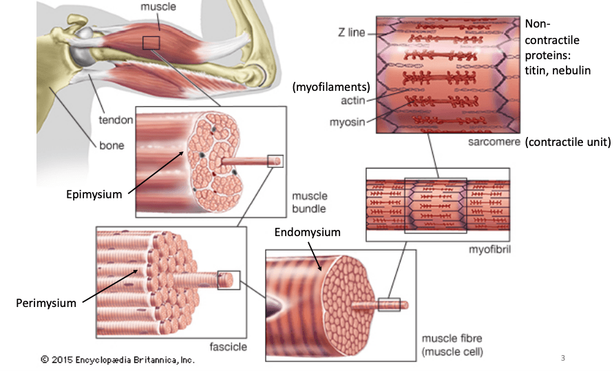
Figure 1: Skeletal muscle structure through different layers of the muscle. Myofascial tissue lies over the epimysium connective tissue which coats the muscle bundle. The epimysium, perimysium, and endomysium are specialized versions of myofascial tissue.

Different forms of fascia can be found all over the body, from encasing organs, to blood vessels and nerves, to muscle.  Fascial tissue that specifically covers muscle, or myofascial tissue, is a thin, white/transparent connective tissue that covers muscle, bundles, muscle fibers, and the muscle as a whole.  If you have ever picked off the thin white stuff covering parts of a chicken breast while preparing it for dinner, you tore off the myofascial tissue layer. Myofascial tissue is an extremely flexible and strong material, which is made up of elastin fibers, for stretch, and collagen fibers, for strength, that are embedded in a gelatinous ground substance, which reduces friction between the muscle fibers and promotes ease of motion [1]. Considered a “deep fascia,” myofascial tissue is made up of a more compacted weave than other fascia found throughout the body and can modify itself depending on the forces placed on it.   (Figure 2).  Because of this, if there is “trauma” or “injury” to the tissue, it can become out of alignment and

Figure 2: 3D visualization of myofascial tissue (white web like structure) and fascia tissue between the skin and muscle (yellow web like structure). Myofascial tissue can be related to a cotton candy structure that is extremely complex and strong. Retrieved from https://www.myofascialrelease.com/about/definition.aspx

cause trigger or dysfunctional points. These points, most commonly referred to as knots, is when the fibers that make up the tissue gets stuck together, loses its elasticity, and becomes taught [2]. Polly de Mille, R.N., C.S.C.S., director of performance services at the Hospital for Special Surgery in New York City explains in an interview for SELF that it is very similar to getting ice cream in silky smooth hair.  When there are “knots” in the muscle fascia, it limits range of motion, and can trigger immune responses which can ultimately lead to pain and discomfort (cytokines have been shown to cause pain and soreness) [3,4].


Now, what is the best way to heal and prevent muscles from experiencing these knots and discomfort? When talking about getting rid of knots in your body, a massage should be the first thing that comes to mind.  The “hurts so good” mentality of deep massaging muscles to where the patient feels pain and then relief afterwards is a popular desire, though not for everyone. Applying pressure and different forces to the tissue through a massage, or foam roller which we will talk about in a little bit, while moving around the fascia helps to separate and relax the tissue and muscle, allowing it to go back to its natural state.  Effects also include an increase in blood flow, which should help muscles get the proper nutrients to repair. Massaging also releases “feel good” brain chemicals, like endorphins, which basically inhibit pain receptors and overall makes you feel better. A study conducted by Mal-Soon Shin and Yun-Hee Sung induced muscle fatigue on 21 young males and treated 11 of them to massages afterwards while recording surface muscle activation and position of their medial gastrocnemius muscle.  According to their study, massaging increases muscle activation and strength due to a change of structural properties. However, in their discussion, they mention that not all messages are effective, which seems to be a common issue in the argument of whether foam rollers, or self myofascial release in general, works or not [5].

Massages are so great because when another person is working out your muscles, they are not only more accurate in pinpointing the location, but they can also apply more force (remember: collagen is EXTREMELY strong in ratio to its size. Proportionally, it is stronger than steel!).  As great as they are, unless someone at home is a masseuse, it can be costly. Self myofascial release techniques, such as foam rolling, have taken over the exercise world and are now regularly used. Foam rolling is when the user applies pressure to “trigger point” or sore spot before or after a workout by using their body weight to roll against a foam cylinder. Though it feels good, does it actually work?

Research has proven that foam rolling is great for warming up muscles and increasing range of motion and flexibility, but the verdict is still out on decreasing muscle soreness.  Though the mechanism behind foam rolling is not exactly known, there is great evidence that it does work on some type of level, whether it is physical or just simply mental. In a systematic literature review of research on using a foam roller before and after workouts, Scott W. Cheatham identified different scientific articles that were critically appraised with trusted conclusions.  From these articles, he identified five studies on the effects of foam rolling and range of motion before exercising. All of these studies resulted with an increase in stretching or range in motion in test subjects [6]. It is common knowledge to stretch before a workout or game to help “warm up” the muscles so that they’re are more flexible, which helps prevent injury and soreness. It is also speculated that rolling out could create a friction that literally heats up the fascia and muscle, making it more flexible and the typical “loose” feeling [2].  After a workout, however, there is a preconception that rolling out will help with delayed onset muscle soreness and pain in general. In this literature review, Cheatham identifies two different journals that conclude that foam rolling does reduce pain, but since the mechanism behind it is still unknown, how much can we trust? In the same SELF article as mentioned previously, Lewis J. Macgregor, Ph.D., an exercise physiologist and lead author of the University of Stirling confirms foam rolling does help increase blood flow, which in turn promotes muscle recovery, but foam rolling does not actually help with myofascial release. Since the collagen in the fascia is so strong, it is argued using your body weight to roll out is not enough. Instead, the pressure of rolling stimulates nerve receptors, which sends the same “hurts so good” feeling to your brain that is then perceived as loosening up the muscle, when really it is not happening.

Overall, foam rolling and myofascial release is an effective way to warm up your muscles and stretch them out before a workout and to help stimulate more blood flow post workout, just don’t get your hopes up about avoiding soreness! Increasing range of motion and flexibility before a workout is a great step to ease muscle soreness, but foam rolling alone is not the answer.  The verdict is still out on the mechanism behind the effects of myofascial release on a cellular level, but hey, if it feels good, why not!


If you’re interested, here is a video of foam rolling techniques because like anything, it’s not effective if you don’t do it properly.

https://www.youtube.com/watch?v=WCj1dvTwOF0


Questions to consider:

Is it worth it to foam roll or stretch in general before physical activity?

Is myofascial release something to think about daily and not just dealing with exercising?

Do you think foam rolling has a placebo effect or is it both systems (nervous and skeletomuscular) working together?

Do you think foam rolling is just another exercising fad?

Would adding heat be beneficial? Or a waste of time?


Sources:

  1. Shah, S., & Bhalara, A. (2012). Myofascial Release. International Journal of Health Sciences and Research,2(2), 69-77.
  2. Fetters, K. A. (2018, July 21). Here’s What Foam Rolling Is Actually Doing When It Hurts So Good. Retrieved April 12, 2019, from https://www.self.com/story/what-foam-rolling-is-actually-doing-when-it-hurts-so-good
  3. Grosman-Rimon, L., Parkinson, W., Upadhye, S., Clarke, H., Katz, J., Flannery, J., … Kumbhare, D. (2016). Circulating biomarkers in acute myofascial pain: A case-control study. Medicine, 95(37), e4650. doi:10.1097/MD.0000000000004650
  4. Zhang, J. M., & An, J. (2007). Cytokines, inflammation, and pain. International anesthesiology clinics, 45(2), 27–37. doi:10.1097/AIA.0b013e318034194e
  5. Shin, M., & Sung, Y. (2015). Effects of Massage on Muscular Strength and Proprioception After Exercise-Induced Muscle Damage. Journal of Strength and Conditioning Research,29(8), 2255-2260. doi:10.1519/jsc.0000000000000688
  6. Cheatham, S. W., Kolber, M. J., Cain, M., & Lee, M. (2015). THE EFFECTS OF SELF-MYOFASCIAL RELEASE USING A FOAM ROLL OR ROLLER MASSAGER ON JOINT RANGE OF MOTION, MUSCLE RECOVERY, AND PERFORMANCE: A SYSTEMATIC REVIEW. International journal of sports physical therapy, 10(6), 827–838.

Isokinetic Dynamometry Engineering Problem

Isokinetic Dynamometry Engineering Problem:

An 18-year-old, 5’4”, 130 lbs female soccer player is just recovering from an ACL tear and wants to know if she can get back in the game. She has been super cautious and tentative to rehab strength exercising routines, so she is sure she is ready, but wants to quantify her strength. To do this, she decides to measure her quadricep and hamstring strength on an isokinetic dynamometer and evaluate her hamstring to quad ratio, which should be between 50 and 80 percent [1]. Other important values include power, work, and peak force to assess the muscle and plan for future rehab exercise routines.

Based on previous studies, to test for power and strength of a muscle, the optimal speed is 60º/sec [2]. She also performs the test with a range of 0-90 º. The results of her hamstring force came back as 54.663 lbs and her recorded quadricep torque value determined by the isokinetic dynamometer is 102.45 lbs*ft3. The patient also wants to know her kick velocity (for fun). The computer is not properly calculating important values, and hand calculations must be performed to ensure accuracy. Calculate the power, work, and force of the quadricep, the angular speed at which the leg kicks, and the hamstring to quadricep ratio to determine if the patient can go back to playing.

*See attached anthropometric weight and measurement documents to determine distance and mass


Solution:
Assumptions: To simplify the math, we are ignoring the effects of gravity and inertia from the swinging limb. In real life, they play a major part in the force read by the machine and there are algorithms the computer goes through to factor out the effects. Also, the math that is performed based on the free body diagram is the forces on the knee joint, not just the quadricep. There are many forces that play a part, but for simplicity reasons we will treat them as only the force produced by the quadricep. Furthermore, there are other considerations when thinking about ACL recovery such as muscle strength and also whether or not the muscles are even. In this problem, we only look at one muscle and do not do a comparison.

1.Since Torque is given (102.45 lbs*ft), Force can be found by

Torque=〖Force〗_quad x d or 〖Force〗_quad*d_perpendicular
First: find perpendicular distance, which will be the length from the patient’s knee to ankle (d on free body diagram)
From the anthropometric table, the distance is Height*(0.285-0.039), where H= 5.33 ft.
Therefore. d=1.312 ft

102.45 lb*ft=〖Force〗_quad*1.312ft
〖Force〗_quad=78.09 lbs

2.Now that Force of the quadricep is found, we can solve for work

The distance, in this case, is the distance the leg travels, which will be the arch distance
s (arch distance)=rθ
r in this case is equal to d. Also, the range (θ) is 90 º, which in radians is 1.571 (90*(π/180))
s=(1.312)(1.571)= 2.06 ft
Now that we have d, multiply by force to get work
Work=78.09 lbs*2.06 ft=160.96 ft*lbs

3.Power can be solved by using the equation

Power=torque*angular velocity
Angular velocity is 60º/sec, which in radians (multiplied by (π/180)) is 1.047 s-1
Plugging in,
Power=102.45lb*ft*1.047 s^(-1)=107.28 (ft*lb)/sec

4.To find angular velocity (ω) her leg is going, the angular acceleration must be determined based off of the force exerted by the quadricep

The following equations must be used
F=m_lowerleg*a_n
a_n=ω^2 r,where r=d
*Note: Since there is a circular motion, angular acceleration must be assessed. Since there is a constant velocity, there is no at component.

Using the weight chart, you should find the weight of the lower leg is 0.0618*weight, which in this case is

(0.0618*130 lbs)/(32.2 )=0.2495 s

Therefore:

78.09 lbs=0.2495 slugs* ω^2*1.047 ft
ω^2=298.7 rads/sec
ω=17.28 rads/sec (times 180/π to get degrees)
ω= 990.2 º/sec

5.Finally, to find hamstring to quadricep ratio

Ratio=(Hamstring Force)/(Quadricep Force) x100
Ratio=54.663/78.09 x100=70%
Therefore, it can be concluded the patient can return back to the field

Overall
Force (quad) = 78.09 lbs
Work=160.96 ft*lbs
Power=107.3 ft*lbs/sec
Angular Velocity = 990.2 º/sec
Hamstring/Quad ratio = 70%
All of these values make physiological sense and line up with average results from other research[3,4]

Tables

Figure 1: Mean Segment data taken from https://exrx.net/Kinesiology/Segments


Figure 2: Anthropometric data showing body segment length as a function of total height. From Winter, D.A., Biomechanics and Motor Control of Human Movement, Wiley Interscience, New York, 1990

Sources:

  1. Rosene, J., Fogarty, T., & Mahaffey, B. (201). Isokinetic Hamstrings:Quadriceps Ratios in Intercollegiate Athletes. Journal of Athletic Training,36(4), 378-383. Retrieved from https://www.ncbi.nlm.nih.gov/pmc/articles/PMC155432/pdf/attr_36_04_0378.pdf.
  2. Duarte, J. P., Valente-Dos-Santos, J., Coelho-E-Silva, M. J., Couto, P., Costa, D., Martinho, D., … Gonçalves, R. S. (2018). Reproducibility of isokinetic strength assessment of knee muscle actions in adult athletes: Torques and antagonist-agonist ratios derived at the same angle position. PloS one13(8), e0202261. doi:10.1371/journal.pone.0202261
  3. Holmes, & Alderink. (1984). Isokinetic Strength Characteristics of the Quadriceps Femoris and Hamstring Muscles in High School Students. Physical Therapy,64, 914-918. Retrieved from http://citeseerx.ist.psu.edu/viewdoc/download?doi=10.1.1.1016.6759&rep=rep1&type=pdf
  4. Heyward, V.H. (2010) Advanced fitness assessment and exercise prescription (6th Ed.) Champaign IL: Human Kinetics


How to Quantify “Getting Back in the Game”

Sports injuries are a major roadblock for athletes, keeping them from playing their best, or even at all. Even after an injury is healed, athletes have to build back up muscles that atrophied over recovery time.  During rehab time, patients undergo different exercise routines to build the muscle that was left unused while the injury was healing. But how do the trainers know when it is safe to allow the athlete back in the field?

One way to quantify the “readiness” of the player is by using an Isokinetic Dynamometer machine to determine how much power the questioned muscle can exert and how that compares to its counterpart.  For example, let’s say a female athlete tears her right ACL. Throughout her rehabilitation, her trainers will set her up on an Isokinetic Dynamometer machine (Figure 1) to determine the power exerted by the right quadricep and hamstring, which are muscles that commonly experience atrophy during ACL recovery, and compare it to the power of her left quadricep and hamstring. Based on the presettings of range of motion, force, and speed, the device can calculate the torque provided by the athlete and then multiply it by the constant speed (isokinetic part) to find the power exerted by those specific muscles. It is obvious that time is needed to heal, but every patient is unique and time could vary. It is important to find a quantitative way to determine when each patient is back to normal strength and this design does just that.

One patent of an isokinetic dynamometer is the “Exercise Physical Rehabilitation and Testing Method and Apparatus” (Patent number: 5,722,937), which was filed in April 17, 1996 and issued on March 3, 1998.  Invented by James F. Smith, it is still used by assignee Cybex International, Inc to this day.  U.S. classifications are as followed: 601/23; 601/24; 482/4; 482/137; 482/142; 482/908.

Figure 1: Set-up of limb to lever arm and dynamometer. The user pushes leg up and back and the dynamometer, comprised of the motor and cycloidal speed reducer, monitors and alerts the computer if the motor needs to slow down or speed up to keep a constant speed depending on the torque exerted by the user.

The main claims of a total 30 for this patent is to help athletes and patients in rehabilitation for muscle atrophy or decreased muscle strength by evaluating the strength of a targeted muscle by forcing the patient to keep a constant speed through resistance. This machine consists of a base with a track to allow adjustability of the chair to customize the fit for each user. There is also a lever arm, where the user will push against during exercises, connected to the chair and to the dynamometer. The dynamometer is comprised of a motor to change torque and a cycloidal speed reducer with a high and low speed shaft to keep a constant speed during exercise (Figure 1).  This machine helps build muscle fibers by forcing the patient/athlete to provide maximum force to move a lever arm while the machine provides resistance (or takes away) to keep the patient moving at a constant speed. Not only can this machine provide biofeedback on the power of the muscle to help physical therapists plan an exercise regimen to help patients, but it can also help athletes build their muscles and ensure their body is balanced to avoid straining and injury. This device has various protocols that subjects the muscles of the user to “concentric or eccentric motion in isotonic or isokinetic modes or continuous passive motion.”

Physical Therapists, Athletic Trainers and athletes will primarily use this machine.  It is very bulky and expensive, so only established facilities can afford this technology.  This product helps physical therapists and athletic trainers assess the muscle strength of their patients to determine what the state of the targeted muscle strength is and help them prepare an exercise routine to get their patients to where they need to be to be healthy and avoid further injury. Athletes can also use this technology to grow muscles due to the max force and full range of motion the lever and program provide.  This product is designed for determining when muscles have developed enough to start playing again after suffering from atrophy during rehabilitation.

Figure 2: Schematic of Isokinetic Concentric mode feedback loop depending on the performance of the user.  If threshold torque is too high, the motor will accelerate. If threshold torque is too low, motor will decelerate to zero speed.

Patients sit in an upright position and strapped at the waist and thigh to stabilize the body and to force the patient to only use the targeted muscle. Next, after setting up the machine with the desired weight and speed, the patient must push and pull a lever arm as hard as they can.  The lever arm, attached to a low speed shaft of a cycloidal speed reducer follows a negative feedback and the machine. For example, Isokinetic Concentric mode, the mode most commonly used for determining the power produced by the muscle, uses the dynamometer control board to determine the angle (boundaries of range of motion) of the lever arm and signals the dynamometer to slow down to a stop until the user pushes the lever arm in the other direction. The torque on the dynamometer control board, which is measured by strain gauges, is sampled every two milliseconds.  The computer monitors the the measured torque (force of the limb attached to the lever arm multiplied by the distance to the targeted muscle) and compares it to the threshold torque. If the measured is greater than the threshold, the motor will accelerate (less resistance) based on the magnitude of the torque and the direction of the measured torque to approach the isokinetic speed. If the measured torque is not sufficient, the motor will decelerate to zero speed until sufficient torque is met (Figure 2).

Compared to other designs, this patent is less costly, smaller in size, and has less parts. Also, the speed reducer incorporated in this design does not create a high pitch noise that is  disruptive in quiet clinical scenes, which was commonly found in previous designs. As for the infrastructure of the machine, this new design fixes a previous problem of slack resistance during start up, which creates a loose and not smooth feeling for patients (also known as backlash). The slack would allow for additional bending torque on the shafts, which creates that loose and unnatural feel. This design fixes this problem because the cycloidal speed reducer specific to this design has a higher torsional stiffness. The cycloidal speed reduces also has a longer life and will reduce the overall effect of backlash throughout time. This patent is still the primary patent for CSMi Medical Solution’s HumacNorm Testing and Rehabilitation System, so it is reasonable to assume these claims are valid and the design is reliable and effective!


Source:

Smith, J.F. (1998). U.S. Patent No. 5,722,937. Retrieved from http://pdfpiw.uspto.gov/.piw?PageNum=0&docid=05722937&IDKey=C45CA52352DA%0D%0A&HomeUrl=http%3A%2F%2Fpatft.uspto.gov%2Fnetacgi%2Fnph-Parser%3FSect1%3DPTO1%2526Sect2%3DHITOFF%2526d%3DPALL%2526p%3D1%2526u%3D%25252Fnetahtml%25252FPTO%25252Fsrchnum.htm%2526r%3D1%2526f%3DG%2526l%3D50%2526s1%3D5%2C722%2C937.PN.%2526OS%3DPN%2F5%2C722%2C937%2526RS%3DPN%2F5%2C722%2C937