Myofascia-nating, does myofacial release work?

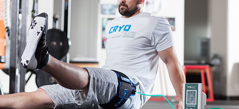
Many people envision an evening under the hands of a masseuse as the perfect example of a relaxing experience, even as those hands dig deep into the “hurts so good” territory. This is often justified with the long term effects promised as the gain to the pain, such as relief of chronic pain, reduced soreness, and feeling looser. These claims are primarily backed by anecdotal evidence rooted in experiences of clients ranging from casual massage goers to professional athletes, as well as from those of practitioners.

Ubiquitously coined by masseuses, physical therapists, and athletic trainers, myofascial release is one of those deep soft tissue massage techniques that gets people excited to let them above their personal pain thresholds. But does myofascial release have any scientific evidence behind it’s effectiveness?

But what really is myofascial release?

The theory is that a primary factor in muscle tightness and pain is the condition of the sticky, gooey web that holds our muscle fibers together and to other parts of our body.

Fig 1: Fascia around and between layers of muscle

Originally posed by physician Stephen Typaldos came the idea that virtually all musculoskeletal injuries were due to distortions of connective tissue, particularly when sticky masses of fascia clump in between muscular fibers. Myofascial release is the practice of, in theory, releasing these fascial clumps to relieve tension on the musculoskeletal system. Dr. Typaldos found a lot of success in his career by using rubbing, friction, sliding, and pulling on acute pain- success that has inspired practitioners to adopt his strategies and aim for the best results.  However, while clumps of fascia melting away is easy to visualize, there is no scientific proof that distortions actually exists, nor that they can be removed by manual work. Some argue that fascia doesn’t even matter while others swear by it and pose theories to back their claims, but currently the mechanism, if any, behind myofascial release is unknown.

There is also no common consensus over how myofascial release is really used. For example, some professionals apply myofascial release to trigger points, which don’t seem to be related to fascia at all as per the video below:

Of course, for anyone looking for relief from pain and soreness myofascial release sounds like a good idea, but the costs of repetitively visiting therapists and masseuses is a deterrent. Thus enters the market of self myofascial release including products such as rollers and massage tools, where one can supposedly achieve myofascial release at home without the need of a practitioner. However, things get a little murky here as well – some argue that these techniques can’t actually achieve myofascial release, and there is no proof in either direction:

https://www.youtube.com/watch?v=BnYdzaoMyQ8

Ultimately, these grievances are rooted in the lack of understanding over the mechanism behind myofascial release. Even as these techniques aren’t tightly defined, we’re still left wondering the question: “But does it work?”

Does the massage work?

This one is tricky to answer, using scientific evidence, as there is a lack of high quality, highly controlled studies I myofascial release massage. A 2013 review on the effect of myofascial release on adults with orthopedic conditions found only 10 peer reviewed articles on the topic. Of these, 6 were case studies of which 5 had degrees of improvement ranging from slight to full recovery, and one on which the treatment failed. Of course, there is no way of knowing if massage really had any effect on those results. Of the studies, it was found that in treating plantar fasciitis, hamstring tightness, and misaligned pelvis myofascial release was useful (especially in plantar fasciitis, with on average 60% better pain reduction than the group without three months down the line!. However, the study with the largest subject was on low back pain, in which it was found that myofascial release did improve back pain, but no better than other manipulation techniques. There was no control in this study, however, so again it’s hard to tell if time alone healed all the subjects. With only one randomized control study (plantar fasciitis), I concurred with the authors here that there was a greater need for stronger studies on the subject.

A 2017 review looked only at randomized controlled trials where individuals and personnel were blinded to which treatment group they belonged in. Only 8 were found, all of which indicated myofascial release was beneficial. The conditions studied were tennis elbow and low back pain. Among these, two of them found that myofascial release on top of physical therapy was more useful than just physical therapy.

As we can see here, there really aren’t many conclusive studies on the matter, without a big enough sample of studies to draw a consensus from. I’ll agree it looks like from what we’ve seen myofascial release therapy seems to help, but only two of the above studies actually compare it to fake massage or other massage techniques. While those two studies found myofascial release was better than faking a massage, two are hardly enough to conclude that myofascial release is responsible for reduced pain and not just any massage. Looking at a third study, we see that there is no significant difference between myofascial release and Swedish massage in pain symptoms.

Okay…but what about foam rollers?

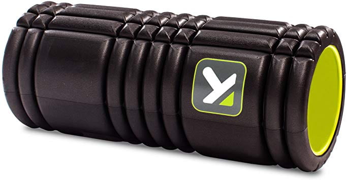
An example of a foam roller

Many people use foam rollers as a cheaper alternative to hands on massage to achieve myofascial release. It doesn’t look like we’re sure if myofascial release is even a real thing, but let’s not just go throwing our rollers out feeling dejected and lied to all along. Even if we aren’t sure how foam rollers work, they may still help.

One study looked at 20 gym-going males and how foam rolling affected them while doing a resistance squat and jump height protocol. They were evenly and randomly split into a foam rolling group and non foam rolling group. The study found that throughout their training which consistent of five consecutive days of exercise, the group that foam rolled had consistently lowered muscle soreness and improved range of motion at each time point. The participants that foam rolled did not have better gains in their squat one rep max, but did have better jump height improvement in comparison to the control. One limitation of this study is that the control group had no replacement to foam rolling, such as just laying down on foam, after their workouts, so there could have been another factor involved in the difference between the two groups.

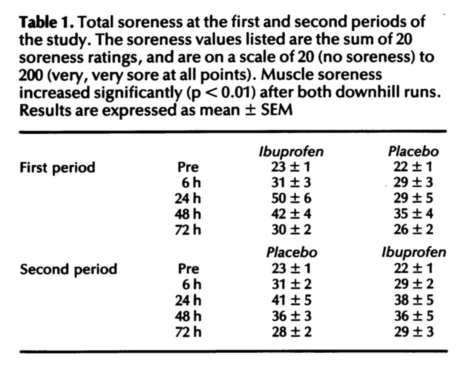
Another study looked at the effect of foam rolling in delayed onset muscle soreness (DOMS), in which a squat regiment was used to induce the pain in both the no foam rolling and foam rolling group, who foam rolled immediately afterward the workout and then 24 hours later and 48 hours later for 20 minutes each time. The foam roller group had significantly reduced muscle soreness and increased tenderness of the quadriceps. The athletes had recorded performance measures such as sprint time and squat reps before the DOMS protocol, and the group the foam rolled had less reduction of performance 24, 48, and 72 hours after. Again, we can say the lack of a more robust control condition applies here, but again the results are promising.

So lets roll it all together

Even though it looks like overall we’re really not too sure what myofascial release massage is, how it works, or if its effective, we can still draw some conclusions from the research. The first is that myofascial release isn’t harmful. With neither the foam rollers or the manual massage did pain increase for subjects or performance decrease. Its true that myofascial release could be no different than any other massage in it’s effects, but they trend to show that whether or not a release of clumps of fascia occurs, the massage does help with pain for certain cases. The same thing goes for foam rolling the legs. Maybe no form of release is occurring at all, but spending the time to foam roll is showing to increase flexibility and reduce soreness at least over the span of time that DOMS is a factor. Importantly, there is no case here to say that if you feel like myofascial release helps you that there is any reason to give it up.


Questions to Consider:

Is it important to know how myofascial release works or just that it does work? If you had limited resources and to support one of those two types of studies, which would it be?

Are randomized controlled trials important to understanding how effective myofascial release is? Or is that being too strict, and looking at case studies and less controlled studies is sufficient enough? Why?

References:

Meltzer, K. R., Cao, T. V., Schad, J. F., King, H., Stoll, S. T., & Standley, P. R. (2010). In vitro modeling of repetitive motion injury and myofascial release. Journal of Bodywork and Movement Therapies, 14(2), 162–171. doi: 10.1016/j.jbmt.2010.01.002

Whitehead, M., Jeffrey, E., khurana, A., Gail, Oster, D., Wilson, S., … Miller, C. (2018, March 8). Self Myofascial Release- What is MFR and how does it work? Retrieved from https://deeprecovery.com/is-myofascial-release-real/

Ingraham, P. (n.d.). Fascia Science Review. Retrieved from https://www.painscience.com/articles/does-fascia-matter.php#sec_origins

Problems MFR Helps. (n.d.). Retrieved from https://www.myofascialrelease.com/about/problems-mfr-helps.aspx

American Fascial Distortion Model Association. (n.d.). Retrieved from https://afdma.com/

Mckenney, K., Elder, A. S., Elder, C., & Hutchins, A. (2013). Myofascial Release as a Treatment for Orthopaedic Conditions: A Systematic Review. Journal of Athletic Training, 48(4), 522–527. doi: 10.4085/1062-6050-48.3.17

Laimi, K., Mäkilä, A., Bärlund, E., Katajapuu, N., Oksanen, A., Seikkula, V., … Saltychev, M. (2017). Effectiveness of myofascial release in treatment of chronic musculoskeletal pain: a systematic review. Clinical Rehabilitation, 32(4), 440–450. doi: 10.1177/0269215517732820

Liptan, G., Mist, S., Wright, C., Arzt, A., & Jones, K. D. (2013). A pilot study of myofascial release therapy compared to Swedish massage in Fibromyalgia. Journal of Bodywork and Movement Therapies, 17(3), 365–370. doi: 10.1016/j.jbmt.2012.11.010

Macdonald, G. Z., Button, D. C., Drinkwater, E. J., & Behm, D. G. (2014). Foam Rolling as a Recovery Tool after an Intense Bout of Physical Activity. Medicine & Science in Sports & Exercise, 46(1), 131–142. doi: 10.1249/mss.0b013e3182a123db

Pearcey, G. E. P., Bradbury-Squires, D. J., Kawamoto, J.-E., Drinkwater, E. J., Behm, D. G., & Button, D. C. (2015). Foam Rolling for Delayed-Onset Muscle Soreness and Recovery of Dynamic Performance Measures. Journal of Athletic Training, 50(1), 5–13. doi: 10.4085/1062-6050-50.1.01

Achey Breaky Muscles

Athletic expertise is not required for one to feel the aching and lasting effects of delayed-onset muscle soreness (DOMS). The aftermath of these effects can often exist along a spectrum ranging from significant muscle tenderness to debilitating pain.¹ In addition, the duration of these effects can exist along a spectrum ranging from 24 to 72 hours.¹ A common misconception correlates a good workout with the subsequent symptoms of DOMS, however, that is just not the case. With that being said, DOMS will often occur in accord with performing new exercises or increasing the intensity and/or duration of a current exercise.¹


The lingering soreness and stiffness as a result of DOMS can often cause individuals to seek methods that will ultimately reduce these symptoms and accelerate their return to the gym. Unfortunately, many of the treatment options on the market have mixed outcomes due to the individualized nature of exercise. Some may obtain adequate results through methods, such as massage or ibuprofen, while others may not; whereas, some methods prove to be completely ineffective, such as cryotherapy and stretching.¹ Furthermore, let’s give a look at a newly proposed treatment for DOMS and whether its results are promising.


Blood-flow restriction (BFR) training is often used at physical therapy as a passive way to regain strength without adding heavy weight. The venous blood supply is terminated at the area of injury, which in turn reduces the amount of oxygen at the site, activates anaerobic metabolism, and encourages muscle hypertrophy.² This proves to be an effective technique for restrengthening the muscle of patients who strictly lift lighter loads.² But, where is the crossover between BFR and DOMS?

Figure 1. BFR machine.[8]

BFR is thought to provide an alternative training method that will achieve similar muscle gains to resistance training with heavy loads while also minimizing the effects of DOMS. It has been postulated that BFR training may attenuate DOMS by preventing calcium-mediated proteolysis³ and by recruiting fast-twitch motor units.⁴ However, it has also been hypothesized that BFR training may induce muscle damage through ischemia-reperfusion (lack of oxygen in the tissues) or through the reduction of neutrophils that aid in inflammatory response.⁵


A 2019 study wished to examine the effects of BFR training on DOMS by using 25 untrained females and submitting them to isokinetic forearm flexion training, where half performed a bicep curl with a BFR cuff at a load reflecting a 30% eccentric-peak torque and the other half at a load reflecting a 30% concentric-peak torque.⁶ The findings revealed neither eccentric or concentric movements with a BFR cuff resulted in DOMS after the 7 days of training.⁶ These results were inconsistent with other studies that showed an increase in DOMS with concentric exercises.⁶ Is this inconsistency due to the low number of participants, the lack of diversity, or the duration of the experiment being only 7 days?


In contrast, a 2017 study compared the effects of BFR training and resistance training on DOMS by using 17 male participants and assessing their elbow-flexor muscle strength for 7 days in one of four training techniques: heavy load, light load, intermittent high-pressure BFR, and continuous low-pressure BFR.⁷ From this, the researchers found DOMS was significantly greater in BFR training (both intermittent high-pressure and continuous low-pressure) than in resistance training (both in heavy and light load).⁷


Much like the other methods of muscle recovery, BFR training may not be the most effective way to minimize DOMS. Exercise can be highly variable and strongly impacted by genetics, which may be the reason a consistent and reliable technique for treating DOMS has yet to be discovered.


Watch this youtube video to learn more about DOMS.⁸

Questions to consider

  • Based on the study mentioned in this article, do you think their findings are an accurate representation of the effects of BFR training on DOMS?
  • How have your opinions regarding DOMS changed with this post?
  • Did the video help you understand DOMS in an easily digestible way?


References

  1. Cheung K, Hume P, Maxwell L. Delayed onset muscle soreness: treatment strategies and performance factors. Sports Med. 2003; 33(2):145-164. DOI: 10.2165/00007256-200333020-00005
  2. Blood-Flow Restriction Training. American Physical Therapy Association. Website.http://www.apta.org/PatientCare/BloodFlowRestrictionTraining/. Updated May 24, 2019. Accessed February 20, 2020.
  3. Sudo M, Ando S, Poole DC, Kano Y. Blood flow restriction prevents muscle damage but not protein synthesis signaling following eccentric contractions. Physiol Rep. 2015;3(7):e12449.
  4. Loenneke JP, Fahs CA, Wilson JM, Bemben MG. Blood flow restriction: the metabolite/volume threshold theory. Med Hypotheses. 2011;77(5):748-752.
  5. Curty VM, et al. Blood flow restriction attenuates eccentric exercise-induced muscle damage without perceptual and cardiovascular overload. Clin Physiol Funct Imaging. 2017;38(3):468-476.
  6. Hill EC, Housh TJ, Smith CM, Keller JL, Schmidt RJ, Johnson GO. Eccentric and concentric blood flow restriction resistance training on indices of delayed onset muscle soreness in untrained women. Eur J Appl Physiol. 2019;119(10):2363-2373.
  7. Brandner CR, Warmington SA. Delayed onset muscle soreness and perceived exertion after blood flow restriction exercise. Journal of strength and conditioning research. 2017;31(11):3101-3108. doi:10.1519/JSC.0000000000001779
  8. Should You Still Train Sore? Youtube. Website. https://www.youtube.com/watch?v=Ut_4C_5CNbg. Published November 18, 2018. Accessed February 20, 2020.

Personalized Blood Flow Restriction. Owens Recovery Science. ORS. 2020c.

Delayed Onset Muscle Soreness: What We Know and What We Don’t (Emphasis on Don’t)

Ever get that feeling two days after a tough run, or a ride that you knew was just a few miles too long, or your first leg day in months (come on, we’re all guilty of that), where you begin to question whether you will ever walk the same again? Walking down the stairs feels like torture, and your quads feel like they get angrier at you with every step you take? Muscle soreness, more specifically delayed onset muscle soreness (DOMS) is common in athletes of all levels of expertise. It occurs after performing a training activity that is unfamiliar. This could be activities than an athlete has not performed in a few months, activities they’ve never performed before, or even simply an intensity level or duration of exercise that they don’t normally reach, despite performing that exercise regularly. These unfamiliar activities, also known as eccentric training, are known to induce severe muscle soreness characterized by increasing intensity of symptoms beginning as late as 24-48 hours after exercise and lasting for days. The underlying physiological mechanism causing DOMS is still unknown and highly disputed, but at least six hypothesized theories for this mechanism have been proposed: lactic acid, muscle spasm, connective tissue damage, muscle damage, inflammation, and enzyme efflux theories [1]. Currently, there exist therapies that have been experimentally shown to decrease DOMS prevalence, including various hydrotherapies [2] and foam rolling [3], but more effective preventative therapies could probably be developed if the underlying physiological mechanism was identified. In order to better understand this phenomenon and the unfortunate encounters I’m sure we’ve all had with it, we are going to look into some of those proposed mechanisms and try to get some insight on how it works (or doesn’t).

Lactic acid is easy to blame for exercise-related muscle pain because of its high production rates during exercise and its perceived role in muscle fatigue and soreness (which is often highly exaggerated). While lactic acid is a common byproduct of exercise, its role in the development of DOMS is likely insignificant. A study performed in 1983 measuring blood lactic acid concentration before and during two different 45-minute treadmill exercises, one on a level surface and one at a 10% decline, found that DOMS was not prevalent in level-surface runners, even though lactic acid concentration was significantly increased. Conversely, downhill runners saw no significant increases in lactic acid concentrations but experienced significant DOMS [4]. There was clearly no relationship between presence of lactic acid and development of DOMS, and the two in fact appeared to be mutually exclusive, so let’s move on to another of the previously mentioned theories.

The inflammation theory initially seems to have a bit more validity, as the similarities between the acute inflammation response, a response to various types of injury including muscle damage, and DOMS are striking. Both phenomena can be characterized by pain, swelling, and loss of function at the area of interest. The time lines seem to match up as well, as both have been reported to increase in severity for about 48 hours and show signs of healing at 72 hours. The issue with this theory though, is the lack of physiological evidence, which is arguably the most important kind. Studies investigating the relationship between DOMS onset and inflammatory biomarkers, like white blood cells and neutrophils, have often failed to find significant results, leading us to believe that inflammation does not cause DOMS [5]. Another drawback of the inflammation theory is the ineffectiveness of anti-inflammatory drugs in preventing DOMS-related pain. A study done using an anti-inflammatory drug and placebo on athletes undergoing eccentric bicycle exercise found no changes in subjective soreness between drug and placebo groups, suggesting that inflammation is not the source of DOMS pain [6]. We won’t completely remove inflammation from the picture though, as it may play more of a role than it appears.

While inflammation itself is likely not the cause of DOMS pain, inflammatory-related processes may not be completely innocent. Bradykinin, an inflammatory mediator, is believed to play a role in DOMS after a study done in 2010 by Murase et al [7]. This study used a previously established rat model of DOMS to show that injecting a B2 (but not B1) bradykinin receptor antagonist 30 minutes before exercise completely prevented DOMS in those rats. The antagonistic effects of the drug used, HOE 140, only last about an hour in the body, and they found that when injecting it 30 minutes after exercise, it had no effect in preventing DOMS. The results can be seen below.

This suggests that bradykinin released during exercise plays a direct role in the development of DOMS, and that preventing that bradykinin from interacting with the B2 receptor prevents DOMS. The role of bradykinin and the B2 receptor in the development of DOMS is not well understood, but it seems like a step in the right direction to me.

There is too much research out there on DOMS to cover in one lowly blog post. I wanted to debunk the lactic acid theory as lactic acid is often a scapegoat for exercise-related pain that is likely sourced elsewhere. While inflammation and DOMS have many similarities that may lead some to believe that there is a causal relationship there, that is also likely not the case. However, there is definitely evidence of some sort of relationship between the two. Further research into the physiological pathway that leads to DOMS is definitely needed to make any conclusive statements on the issue, and the bradykinin B2 receptor pathway is probably a good place to start. But until then, you’re just going to have to suck it up next time you feel like your quads will never work again two days after your new leg routine. Many have been there and survived before. You will too.


Questions to consider:

What distinguishes DOMS from standard muscle soreness?

Think about any times you may have experienced DOMS- what were you doing and why do you think it led to DOMS?

How could you determine the presence of DOMS in animal models when it cannot be subjectively reported? (Hint: check reference 7 for ideas)

How could preventative therapies for DOMS promote better health and wellness?


References:

[1] Cheung, K., Hume, P. A., & Maxwell, L. (February 01, 2003). Delayed Onset Muscle Soreness: Treatment Strategies and Performance Factors. Sports Medicine, 33, 2, 145-164.

[2] Vaile, J., Halson, S., Gill, N., & Dawson, B. (March 01, 2008). Effect of hydrotherapy on the signs and symptoms of delayed onset muscle soreness. European Journal of Applied Physiology, 102, 4, 447-455.

[3] Pearcey, G. E., Bradbury-Squires, D. J., Kawamoto, J. E., Drinkwater, E. J., Behm, D. G., & Button, D. C. (January 01, 2015). Foam rolling for delayed-onset muscle soreness and recovery of dynamic performance measures. Journal of Athletic Training, 50, 1, 5-13.

[4] Schwane, J. A., Watrous, B. G., Johnson, S. R., & Armstrong, R. B. (January 01, 1983). Is Lactic Acid Related to Delayed-Onset Muscle Soreness?. The Physician and Sportsmedicine, 11, 3, 124-31.

[5] Smith, L. L. (January 01, 1991). Acute inflammation: the underlying mechanism in delayed onset muscle soreness?. Medicine and Science in Sports and Exercise, 23, 5, 542-51.

[6] Kuipers, H., Keizer, H. A., Verstappen, F. T., & Costill, D. L. (January 01, 1985). Influence of a prostaglandin-inhibiting drug on muscle soreness after eccentric work. International Journal of Sports Medicine, 6, 6, 336-9.

[7] Murase, S., Terazawa, E., Queme, F., Ota, H., Matsuda, T., Hirate, K., Kozaki, Y., … Mizumura, K. (January 01, 2010). Bradykinin and nerve growth factor play pivotal roles in muscular mechanical hyperalgesia after exercise (delayed-onset muscle soreness). The Journal of Neuroscience : the Official Journal of the Society for Neuroscience, 30, 10, 3752-61.

DOMS: Why do your muscles hurt days after exercise?

Chris Hernandez and Christian Poindexter

Soreness is a typical and often expected side effect of any moderate level of physical activity or exercise.  However, contrary to popular belief, there are many different types of soreness which are a result of separate things. For example, the soreness that many people experience during or immediately after exercise is known as acute soreness. Acute soreness typically develops within a couple of minutes of the muscle contraction and dissipates within anywhere from a few minutes to several hours after the contractions have ended[1].  It is widely accepted that this soreness is a result of the accumulation of chemical byproducts, tissue edema, or muscle fatigue.  Delayed Onset Muscle Soreness (DOMS) typically develops between 12-24 hours after muscle contractions end, with peak ‘soreness’ being experienced 24-72 hours after the exercise is over[1].  Exercises typically associated with DOMS include strength training exercise, jogging, walking down hills, jumping, and step aerobics. Apart from soreness, people suffering from DOMS also experience swelling in their sore limbs, stiffness of adjacent joints, tenderness to the touch, and temporary reduction of strength in affected muscle[1].  Unlike with acute soreness, there are several competing theories on the cause of DOMS, none of which have been ultimately proven to be the predominant cause.  

One of the first and most touted theories was the Lactic Acid Theory.  This was based on the concept that the muscles continue to produce and accumulate lactic acid even after the exercise is abated.  The accumulation of this lactic acid is thought to cause the noxious stimulus associated with soreness[2].  The paper we are using, “Delayed Onset Muscle Soreness: Treatment Strategies and Performance Factors”, cited a study done by French researchers regarding misconceptions about lactic acid, and more specifically lactate[3].   This study goes on to explain that during the recovery phase post-contraction, accumulated lactate gets oxidized by lactate dehydrogenase (LDH) into pyruvate.  This pyruvate is either oxidized in the mitochondria where it contributes to the resynthesis of ATP, or it is transported in the blood to be used or disposed of elsewhere in the body[3].  It has been observed that for test subjects whose lactate levels were monitored for 72 hours before, during, and after exercise, their lactic acid levels returned to pre-exercise levels within 1 hour of the cessation of exercise[2].  Since DOMS does not set in for 24-48 hours, it is very unlikely that lactic acid accumulation is the cause of the pain and other symptoms associated with this disease.  

These researchers did note some conditions however that were noted to affect the lactate levels of those participating in the study.  For example, a participant with a diet rich or low in carbohydrate concentrations can cause lactate levels to decrease or increase respectively[3]. Further, participants who had undergone strenuous exercise the day before are likely to show signs of glycogenic depletion, which could cause them to have irregular lactate levels[3]. Further, the type of exercise performed was also shown to have an effect on not only lactate levels but also on the time frame required for levels to return to normal[3].  To improve this study and potentially get better results it would be best to make sure that all test subjects were undergoing the same exercise regimens.  It would also be beneficial if the amount of carbohydrates (based on body weight) was held standard, and that they all experienced 48 hours of rest before data collection[3].  However, even given these potential weaknesses, given that the lactate levels return to below normal within an hour of exercise cessation, it can be said with reasonable certainty that lactic acid is not the cause of DOMS[2,3].

A more current and well-supported theory is the Muscle Damage theory, which is based on the disruption of the contractile component of muscle tissue after eccentric exercise. Type II fibers have the narrowest z-lines and are particularly susceptible to this type of disruption. Nociceptors located in the muscle connective tissue and in the surrounding tissues are stimulated, which leads to the sensation of pain that we know as DOMS. In practice, muscle soluble enzymes can be used as an indicator of z-line disruption and sarcolemma damage. Creatine Kinase (CK) is used as one of these muscle permeability indicators; any disruption of the z-lines and damage to the sarcolemma will enable the diffusion of  CK into the interstitial fluid, where it can be measured.

To test the connection between eccentric exercise and changes in CK, this study[4] used five healthy adults and had them walk on a treadmill for an hour at a 13-degree incline to test the effect of concentric exercise, and then a 13-degree decline five weeks later to test eccentric exercise. Venous blood samples were taken pre-test and every 24 hours until CK levels had returned to pre-exercise levels. Following downhill walking, all subjects reported muscle pain and tenderness in the calves and glutei muscles, which developed several hours post-exercise and was maximal between 1-2 days after. The severity of pain differed between subjects, but following uphill walking none reported any pain or tenderness. Both concentric and eccentric exercises showed increases in CK levels, but eccentric work showed much greater levels and peaked after 4-7 post exercise, as shown in figures 1-2.

Newham, et al. conclude that rises in CK levels are a result of eccentric work, and suggests that the extent to which muscle is lengthened and the level of habituation to eccentric work play a role in the enzyme response, and thereby in DOMS. However, they acknowledge that there is only a correlation between CK levels and DOMS and not necessarily a causation. Additionally, the sample size they used is small which could lead to inaccurate data.

There are many theories of the cause of DOMS, and none can fully explain the phenomenon. A combination of models has also been proposed[5], drawing aspects from various theories. Of the two theories we examined, the muscle damage theory was the most conclusive, showing a relationship between plasma CK levels and DOMS. Additional studies will be necessary to determine whether the muscle damage theory, another theory or possibly a combination of multiple can best explain the symptoms of DOMS.


Questions to Consider:

  1. Which types of exercise would help to prevent DOMS?
  2. How would you better design an experiment to correlate DOMS with enzyme activity?
  3. Can we conclusively rule out lactic acid as an explanation?


References/Further Reading:

[1] American College of Sports Medicine. Delayed Onset Muscle Soreness. Delayed Onset Muscle Soreness, American College of Sports Medicine, 2011, www.acsm.org/docs/brochures/delayed-onset-muscle-soreness-(doms).pdf.

[2] Schwane JA, Hatrous BG, Johnson SR, et al. Is lactic acid 63. Hasson SM, Wible CL, Reich M, et al. Dexamethasone related to delayed-onset muscle soreness? Phys Sports Med phoresis: effect on delayed muscle soreness and muscle function 1983; 11 (3): 124-7, 130-1

[3]Léger , L., Cazorla , G., Petibois , C. & Bosquet , L. (2001). Lactate and exercise: myths and realities. Staps , n o 54, (1), 63-76. doi: 10.3917 / sta.054.0063.

[4]Newham, D. J., Jones, D. A., & Edwards, R. H. T. (January 01, 1986). Plasma creatine kinase changes after eccentric and concentric contractions. Muscle & Nerve, 9, 1, 59-63.

[5]Cheung, K., Hume, P. A., & Maxwell, L. (2003). Delayed Onset Muscle Soreness. Sports Medicine,33(2), 145-164. doi:10.2165/00007256-200333020-00005

[6]Armstrong, R. B. (January 01, 1990). Initial events in exercise-induced muscular injury. Medicine and Science in Sports and Exercise, 22, 4, 429-35.

No Pain, No Gain: Stop taking those NSAIDs!

Most athletes have heard the term, “no pain, no gain” at one point or another in their athletic careers, but this saying is truer than one might think. Having grown up in a household where sports were played year round, it was common to take over the counter non-steroidal anti-inflammatory drugs (OTC NSAIDs), such as ibuprofen or aspirin to ease the pain. Ran too far? Easy solution, take some Advil. Overdo it with the lifting? Take a few Motrin and you’ll be ready in the morning. It was even common to take these over the counter drugs before a workout, as a way to get a head start on the pain.

Ibuprofen, a common OTC NSAID.

Is there any validity to this solution? OTC NSAID’s are known for their ability to reduce fevers and minor aches and pains. With that being said, are they really useful for exercise related injuries or pains? Specifically, are they safe and effective to use for delayed onset muscle soreness (DOMS) due to exercise?

DOMS is the pain and stiffness that is felt typically between 24 and 72 hours after the workout has been completed but can last up to 7 days. After a strenuous workout, the body responds with acute inflammation as a natural way to heal the body. This inflammation usually goes hand in hand with DOMS, but the specifics of this relationship have not been clearly defined. Many athletes try to combat this by taking ibuprofen (or other OTC NSAIDs) to ease the pain, but recent studies have shown that this isn’t necessarily a foolproof plan.

In a 2006 study completed by Nieman et al., the influence of ibuprofen was measured during the 160-km Western States Endurance Run on endotoxemia (the presence of endotoxins in the blood), inflammation and plasma cytokines. The study included 29 ultramarathoners who consumed 600 and 1200mg ibuprofen the day before and on the race day, respectively, and 25 controls that competed in the race but did not take ibuprofen or any other medications. Blood and urine samples were collected the morning prior to and immediately following the race, and subjects recorded muscle soreness during the week following the race using a 10-point Likert scale. It was found that ibuprofen use compared to non-users did not alter muscle soreness or damage. In addition ibuprofen use was linked to elevated indicators of endotoxemia and inflammation. One limitation of the study was that it did not have a placebo group due to ethical concerns from the race director, but they may have had an impact on the results. In addition, race conditions are not the best conditions to conduct an experiment under, as they can cause extra stress on the body, affecting the results.

In another study conducted by Donnelly et al., 32 volunteers participated in a study in which ibuprofen was tested against an identical placebo for its effectiveness in reducing muscle soreness and damage after two periods of downhill running. Volunteers took two 600mg ibuprofen or placebo tablets 30 minutes before each run, and took one 600mg tablet every six hours up to 72 hours post-exercise. Blood samples were drawn pre- and post-exercise, and at 6, 24, 48 and 72 hours and analyzed for indicators of muscle damage and inflammation. A questionnaire was used to determine muscle soreness for different regions of the body (the results can be seen in Table 1). The results indicated that ibuprofen is not an appropriate treatment for DOMS and muscle damage. However, one limitation of this study was that during the 10 week break between the two periods of running, there was no monitoring or control of the participants, which means their lifestyles could have been very different from each other, thereby affecting the results.

Furthermore, it is known that exhaustive physical activity leads to small intestinal injury and short-term loss of gut barrier function in otherwise healthy individuals. Another study, conducted in 2012 reveals that ibuprofen aggravates this exercise-induced small intestinal injury and induces gut barrier dysfunction in healthy individuals.

Based on this research, I have concluded that OTC NSAIDs should be discouraged as a way to mitigate the pain that comes with delayed onset muscle soreness. The data from the first two studies show that ibuprofen (and implied other NSAIDs) are not a satisfactory way to decrease the pain that comes from delayed onset muscle soreness. The third study shows that ibuprofen can be extremely harmful to the user and therefore should not be taken if it can be avoided. Based on this information, not only should OTC NSAIDs not be taken for DOMS, but the relationship between inflammation and DOMS should be more thoroughly investigated. It could be that the inflammation isn’t causing DOMS and that is why the drugs aren’t relieving the pain. However, the first study suggests that the intake of ibuprofen increased inflammation, the exact opposite of what it was supposed to do. Either way, ibuprofen and other NSAIDs should not be taken for relief from DOMS.

Questions to consider:

Do you take OTC NSAIDs such as ibuprofen or Advil when you are feeling sore after a hard workout? Do they help? Will you continue?

How often do you think people who exercise regularly take OTC NSAIDs? Should this change?

What may be an alternative to taking NSAIDs for muscle soreness?

Do you still think it is safe to take NSAIDs for other types of pain, such as menstrual cramps, headaches or fevers?

Further Reading:

An article looking at the prevalence of using analgesics (includes NSAIDs) in exercise – related pain

An article looking at the effect ibuprofen has on neutrophils (white blood cells that are an important part of the inflammatory response)

Last year’s blog post discussing at delayed recovery after exercise due to NSAIDs