Skip to content

About cmanneri

Christy is a graduate of the Biden School of Public Policy and Administration at the University of Delaware with a master's in Public Administration. She is the chief operating officer of a local non-profit, 3B Brae’s Brown Bags and a Communications Specialist for the University of Delaware. Her research interests include issues of social justice, such as nutritional insecurity, community re-entry, and domestic violence. She also volunteers with her local school district and has a deep passion for education policy, as she believes many social injustices stem from inequitable opportunities in education.

Can your laboratory section frozen specimens?

We do not currently have the equipment to produce frozen sections, though we are considering addition of this service in the near future.  In the meantime, cryostat sectioning is available through the Delaware Biotechnology Institute (DBI) Imaging Core Facility and the A.I. duPont Children’s Hospital Histology Laboratory.

Can you perform additional stains beyond H&E?

At present, our laboratory only performs special stains for disease organisms commonly encountered in diagnostic service (Gram stain, GMS, etc.).  We can advise laboratories on other stains which may be applicable for a specific area of interest and can order and perform these additional staining procedures upon request.  Please contact the pathologist for more information.

Do I have to submit tissues in cassettes?

We welcome submission of fixative containers with either pre-trimmed tissues in cassettes or untrimmed tissues.  Feel free to contact the pathologist if you require assistance with tissue collection and handling or proper orientation in cassettes.  Trimming your own tissues is also a way to save on service fees and cassettes can be provided by our laboratory for a nominal cost.  However, tissues do not have to be submitted in cassettes, and our histotechnician routinely trims specimens submitted to our service.  Specimens MUST be submitted in an adequate volume of fixative (if unsure what to use, see “Choosing a Fixative”) regardless of whether they are whole or pre-trimmed in a cassette.  Containers with formalin must have appropriate formalin-hazard labels.  For further information, please read “Submitting a sample”

Can I cut and embed my own tissues using your equipment?

We can also train users on proper embedding techniques upon request.  Trained users can then self-embed tissues at our embedding station upon arrangement with the histotechnician.  Due to liability issues and potential costs related to service and repair following non-technician use, we cannot currently allow clients to perform microtomy (sectioning) using our equipment.

What information should I provide to the pathologist reviewing slides?

Just as you would not want your doctor to attempt diagnosis of an illness without hearing about your symptoms and performing a physical examination, the pathologist needs to be informed of important clinical and specimen information before microscopic analysis.  This information includes:

  • Animal information- species, breed, sex and castration status, age
  • Clinical information- duration and signs of disease, results of other diagnostic testing
  • Specimen information- the organ or tissue and specific anatomic location of each specimen, how the specimen was collected, and gross lesions observed in the specimen.  For lesions, please describe: size (measure if possible), shape, color, consistency, distribution.

NOTE: The pathologist typically does not observe the gross specimen prior to tissue processing and sectioning.  It is imperative to describe what you saw in the body and specimen prior to trimming and fixation.

Clinical reasoning- What insight are you hoping to gain from microscopic tissue analysis?  What lesions are you expecting the pathologist observe (for experimental disease agent exposures)?  What is/are your working diagnosis or differential diagnoses?

Research protocol- experimental groups, known disease exposure, group treatments and time course, etc. (for research cases only).  Additional protocol sheets can be attached to the submission form as needed.

A summary of the information listed above should be provided with the specimen accession form [Link to Accession Form].  Printed protocols and other supplemental information (digital photos, diagnostic test reports, etc.) can be attached to the accession form if necessary.


When can I expect my finished slides or blocks?

This is a brief overview of services offered by our laboratory.  Please see accompanying  fee schedule for current service rates.

Trimming

Tissues received in fixative will be manually trimmed by an ASCP-certified histotechnician to meet the size and orientation specifications required for processing and embedding.  Clients have the option of performing this step prior to submission to reduce preparation fees.  The Comparative Pathology Laboratory can provide training for proper sample trimming and orientation upon request (see fee schedule).

Processing

Trimmed specimens undergo overnight processing in a Leica processor to dehydrate the specimens in preparation for paraffin embedding.

Embedding

Processed specimens are manually transferred from cassettes to metal wells and are embedded in paraffin wax at a Leica embedding center by the histotechnician to maintain proper specimen orientation for sectioning.  Embedded specimens (“Blocks”) are complete at this step.

Sectioning

Paraffin blocks are affixed to a Leica microtome and typically cut to 4-6 micron thick sections.  Sections are transferred from a water bath to a microscope slide for later staining and histopathologic analysis.  Unstained slides are complete at this step.

Staining

Routine histopathologic analysis utilizes Hematoxylin and Eosin (H&E) staining applied by a Leica autostainer.  Special stains, such as Gram, Periodic Acid Schiff (PAS), and other additional stains, are applied manually by the histotechnician.  Contact the pathologist for special stain availability and cost.  Stained slides are complete at this step.

Decalcification

Bone and other mineralized specimens require chemical decalcification prior to processing to soften tissues for sectioning.  This additional step in specimen preparation creates a delay in processing of one or more days and can also create loss of detail in soft tissues and bone marrow which can interfere with microscopic analysis.  Please contact the pathologist if this may be a concern in your study.

Digital slide scanning and virtual archives

Prepared glass slides are scanned by an Aperio AT2 ScanScope to create virtual slides which can be archived and analyzed indefinitely via Leica Biosystems' eSlide Manager and Image Analysis Toolbox.  Virtual slides are viewable at all magnifications up to that of the objective (20x or 40x) under which scanning was performed.  Archived specimens can be shared with collaborators at the client's discretion.  Once scanned, remote viewing and analysis of archived slides are possible in real time at any location worldwide where internet services are available.

Histopathologic (Microscopic) Analysis and Reporting

Stained slides are analyzed by an ACVP-certified veterinary anatomic pathologist using one of the laboratory’s Leitz or Olympus light microscopes or through Aperio virtual microscopy.  Results generated from the analysis are typically reported as a descriptive report and/or electronic spreadsheet. Reports provide a morphologic diagnosis, i.e. a concise technical description of the pathologic process occurring in the tissue, and/or a description of the lesions observed in the tissues on each slide.  A summary of pertinent lesions by tissue or treatment group and clinical correlation of lesions observed to signs observed in the in the study is also provided when applicable.  Final reports can be mailed or transmitted electronically by email based upon client preference.

Digital Photography

High quality unaltered digital images of microscopic features of interest are captured by an ACVP-certified veterinary anatomic pathologist and provided in .tif or .jpg format based upon client preference and intended use.  Images include descriptions and annotations prepared by the pathologist.

Consultation and TrainingDSC_6610

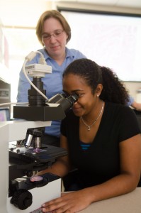
Laboratories utilizing the UD Comparative Pathology Laboratory have unique access to one-on-one or small group consultation and training by a board-certified veterinary anatomic pathologist and registered histotechnician prior to and during use of our services.  Consultation and training topics include, but are not limited to: gross anatomy and lesion recognition; tissue collection, fixation, trimming and embedding; special stain options; microscopic analysis and experimental design.  A brief consultation can be conducted at no charge prior to sample submission to determine individual client needs for services, training, or additional consultation.  Sessions can be conducted by email, phone, or in person based upon client preference.  Training sessions are held in our laboratory (for trimming and embedding) or in the client’s laboratory (for anatomic localization, lesion identification, and sample collection).   Following training, clients may also elect to self-embed tissues using our equipment during normal business hours to reduce the cost of tissue preparation.

Joanne Kramer is an ASCP-certified histotechnician with 6 years of service experience.  She is a graduate of Delaware Technical Community College.


Erin Brannick is a veterinarian and ACVP-certified veterinary anatomic pathologist with experience in necropsy and histopathology in wide variety of animal species (avian, large animal, small animal, zoo/exotic animal, and laboratory animal), mouse phenotyping, and immunohistochemistry.  She is a graduate of The Ohio State University College of Veterinary Medicine.


Dr. Brannick's website: http://ag.udel.edu/anfs/faculty/brannick.html