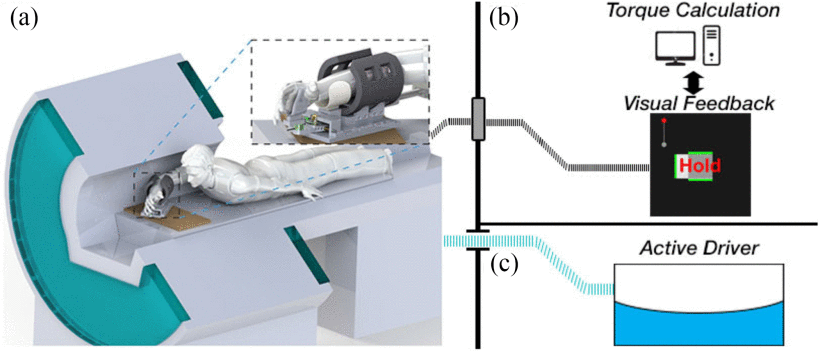
We are interested in understanding the mechanical behavior of individual muscles during coordinated joint motion. Currently, methods such as sEMG and ultrasound elastography can only be used to quantity individual muscle forces in a limited set of superficial muscles. In our research, we use magnetic resonance elastography (MRE) to obtain shear wave velocities from all muscles in the forearm with measurements of joint torque and angles to quantify forces of individual muscles during isometric actions. Our MR-compatible robotic device, the MRE-bot, allows us to safely measure wrist joint torques at different joint postures during magnetic resonance imaging (MRI). Our work focuses on quantifying individual muscle forces in the forearm to evaluate their role in the control of wrist joint tasks. The work on this project is done in collaboration with the Mechanical Neuroimaging Lab. In this protocol, individuals are cued to reach and maintain a desired wrist torque target in either the flexion/extension or radio/ulnar deviation directions. Simultaneously, a pneumatic actor and MRE scan are used to measure shear wave velocities of 3D volume encompassing all forearm muscles.
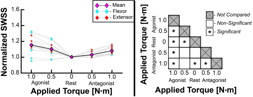
Measurements of shear wave speed squared (SWSS) are normalized using the value of each muscle during the rest condition (torque = 0 Nm). The normalized SWSS values for each muscle are plotted below for both flexors and extensors. Agonist torque is defined during flexion for a flexor and during extension or an extension muscle. A one way ANOVA with post hoc analysis was performed to determine the pairs of contraction states that are significantly different from each other.
Publications on this topic
C. A. Helm, F. Sergi, “Model-Based Estimation of Active and Passive Muscle Forces Using MRE in Forearm Muscles During 2-DOF Wrist Tasks”, doi: 10.1101/2024.02.15.580561, 2024, pre-print.
D. R. Smith, C. A. Helm, A. Zonnino, M. D. J. McGarry, C. L. Johnson, F. Sergi, “Individual Muscle Force Estimation in the Human Forearm Using Multi-Muscle MR Elastography (MM-MRE)”, IEEE Transactions on Biomedical Engineering, vol. 70, no. 11, pp. 3206-3215, Nov. 2023, pre-print, available online.
A. Zonnino, D. R. Smith, P. L. Delgorio, C. L. Johnson, F. Sergi, “MM-MRE: a new technique to quantify individual muscle forces during wrist isometric contractions using MR Elastography”, 16th International Conference on Rehabilitation Robotics, 2019, pre-print, available online.
