Sweat Sensor Channel Geometry

Identify

Soft microfluidics revolutionized the development of sweat sensors by providing increased flexibility so the device can be worn on the skin while an individual is exercising. The material selected for these sensors is important for the flexibility and comfort of these devices. Elastomers with a low modulus but high elasticity are typically chosen as they allow the user to easily move while still obtaining sufficient sweat collection by close contact and flexibility upon the skin [1]. As these types of materials can more easily deform, it is important to consider the geometric design of these microfluidic channels. It is important the sweat sensor can withstand external forces or pressures, such as the individual touching the sensor, especially during exercise. These channels should not easily deform as significant deformation could greatly interfere with the volume of sweat collected, in addition to other measurements like sweat rate and the measured analyte concentrations.

It is important to first understand how externally applied forces influence the deflection of the microfluidic channels. We can then understand how the volume of these channels changes in response to applied forces which will allow us to select the optimal geometry that minimizes the change in volume to increase the accuracy of the device in realistic conditions [2]. If we want to account for externally applied forces that might be applied to the sweat sensor during use, should we design narrow or broad channels? Should there be considerations for height? To understand these questions, we will need to understand how the width and height ratio of microfluidic channels influences the percentage of volume change within these channels.   

Formulate

To solve this problem, we will assume that the applied pressure acts as a uniformly distributed vertical load across the channel width. For simplicity we will only be calculating the results for a single channel, but the principles could be used to calculate the entire surface of the sweat sensor. Since the channels of sweat sensors are often rectangular, we will model the sweat sensor as a rectangular beam that is fixed on both ends. Once the sweat sensor is placed onto the skin we are assuming it is permanently in place as any movement of the sensor would ruin the obtained results. We are also assuming the channel has uniform thickness.

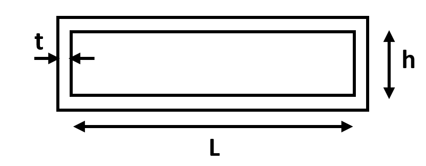
Figure 1. Cross section of microfluidic channel. For our calculations, L will be the width of the channel, h the height of the channel, and t the thickness of the channel. The channel length is a.

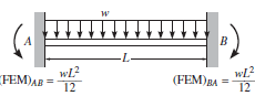
Figure 2. Channel free body diagram. Free body diagram of a beam fixed at both ends with uniform distributed loading applied [3]. L is the width of the channel.

Solve

The free body diagram of Figure 2 can be solved to find the vertical reaction forces at the fixed ends. By symmetry, the two vertical reaction forces are equal to the force of the distributed load which is the load times the length. Each vertical reaction force is equal to wL/2.

We can calculate the bending moment equation by making a cut in the beam to understand internal forces and moments and solve for the bending moment.

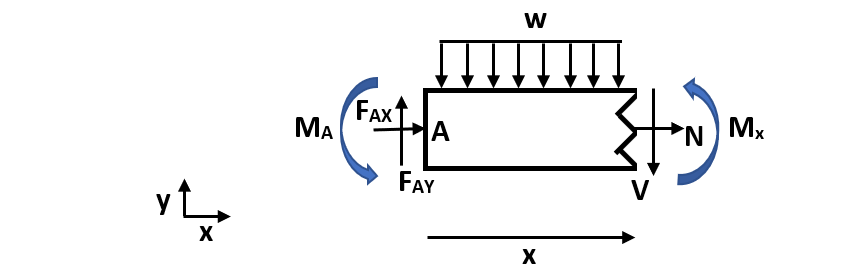
Figure 3. Cut channel section. In this figure, w is the distributed load, N is the normal internal force, V is the internal shear force, Mx is the internal moment at an arbitrary cut, FAX and FAY are reaction forces at fixed end A, and MA is the fixed end moment at A. Mx is defined in the positive direction and x is an arbitrary length along the total width (L) of the beam.

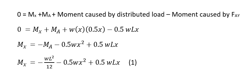
Solve for the bending moment equation:

The sum of the moments equals 0 and any horizontal forces do not create a moment about point A. As shown in Figure 2, the moment about a fixed end for a uniform distributed load is equal to wL2/12. 

Calculate deflection:

Calculate change in volume:

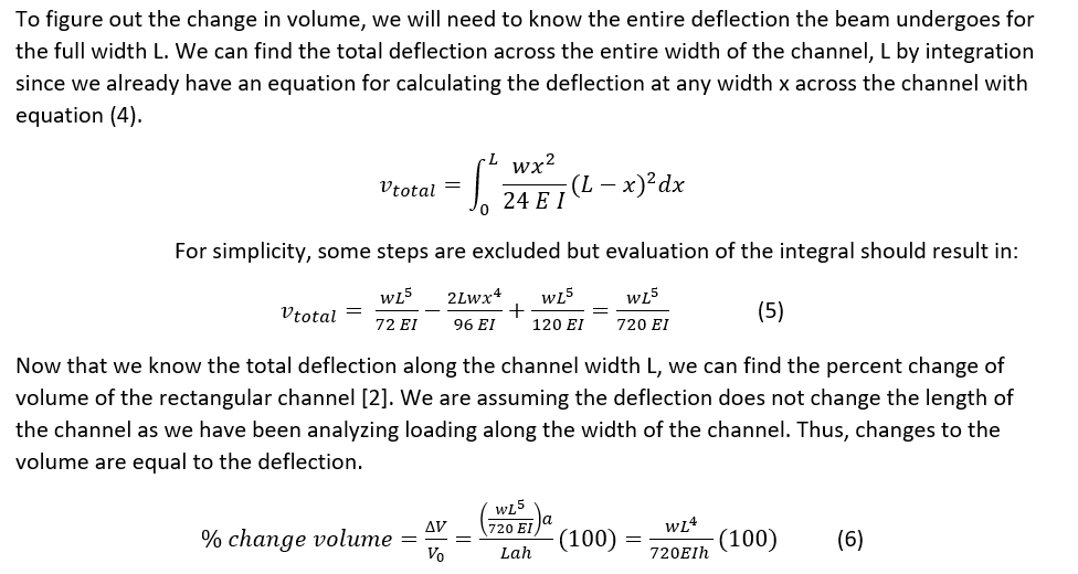
Equation (6) gives us the relationship between the width (L) and the height (h) of the channel so we can understand how the channel geometry influences the change in volume of the channel with externally applied forces. A positive change in volume is reasonable as our deflection of the channel was positive as well. These results were also found in a study analyzing colorimetric sweat sensors that found that narrow channels had a smaller volume change compared to broad channels [1]. There are several limitations to these calculations for practical applications. We analyzed the influence of deflection that caused a change in the height by vertical loading, but realistically the forces might have other components. Depending on how the sweat sensor adheres to the skin, the assumption of fixed ends may not always apply. There may also be instances in which the force applied is not uniformly distributed, but may act at a specific point.

Although the channel height is important to consider, the channel width is a much greater influence on the change in volume of the channel with deflection as it is raised to the fourth power. The height is only raised to the first power which has a less impact on the change in volume when considering external forces. We should aim to use narrow channels in the microfluidic channel design of our sweat sensor compared to broad channels as increasing the width increases the amount of deflection of the channel by equation (5) and produces a greater change in the volume of the channel.


References

[1]. Koh A, Kang D, Xue Y, et al. A soft, wearable microfluidic device for the capture, storage, and colorimetric sensing of sweat. Sci Transl Med. 2016;8(366):366ra165-366ra165. doi:10.1126/scitranslmed.aaf2593

[2]. Rogers, John, et al. Soft, wearable microfluidic systems capable of capture, storage, and sensing of biofluids. United States Patent WO2017218878 A1.  United States Patent and Trademark Office. 16 June 2017.

[3]. Engineering Stack Exchange. Structural engineering – How to determine fixed end moment in beam? https://engineering.stackexchange.com/questions/15040/how-to-determine-fixed-end-moment-in-beam. Accessed May 14, 2020.

To Stretch or Not To Stretch

Stretching is regularly included in exercise regimes by athletic trainers and coaches and is often recommended to novice athletes. The chronic effects of stretching result from consistent practice and are thought to be a preventative measure to reduce risk of injury by increasing flexibility to increase overall range of motion. [1] This increased range of motion is thought to increase overall performance. However, those against stretching argue the long-term effects could lower performance by decreasing muscle strength.

Types of Stretching

Three common types of stretching include static, dynamic, and proprioceptive neuromuscular facilitation (PNF). [2] Static stretching involves lengthening a specific muscle group for a period of time without movement. This method is arguably the safest form of stretching, especially for beginners, as it minimizes the risk of tearing or straining the muscle by overstretching. [3] PNF incorporates static stretching in addition to isometric contraction and relaxation of the muscle. Unlike static and PNF methods, dynamic stretching involves active movements to increase the range of motion while raising the heart rate before an exercise.

Mechanisms to Increasing Flexibility

Several mechanisms describe how stretching can increase flexibility over time including increasing compliance and increasing stretch tolerance. Compliance relates to the elasticity of the muscle-tendon unit and is useful in generating forces as elastic energy is stored by eccentric contractions during the stretch-shortening cycle (SSC). [4] Stretching can increase the compliance of the muscle-tendon, increasing the energy potential. Increasing the stretch tolerance of a muscle is a second mechanism to increase flexibility. Long-term stretching can alter how the central nervous system receives signals from structures aiding in proprioception and regulation of muscle stiffness including nociceptors, Golgi tendon organs, and muscle spindles. [5] Altering these signals may result in greater ranges of motion with decreased resistance by the nervous system.

Stretching Reduces Injury in Some Sports

Research has shown that stretching can reduce injury by increasing flexibility, but only in some sports. For sports requiring jumping motions that involve high intensity SSCs, like soccer and football, stretching has been shown to reduce injury. [4] In these sports, the muscle-tendon system works as an elastic spring. With a compliant unit, potential injury is reduced as greater energy can be absorbed by the tendon, sparing the muscle fibers potential damage. However, if the tendon has low compliance greater forces can be transferred to the muscle, resulting in injury if the muscle is unable to support high amounts of energy.

A prospective study published in 2003 from Ghent University measured initial muscle flexibility for 146 male professional soccer players and analyzed how flexibility related to the development of muscle injuries throughout the season. Goniometers were used to measure the flexibility of the hamstring, quadriceps, adductor, and gastrocnemius muscles on both sides of the athletes. The study reported no statistical significance between players height and weight, but did not analyze other factors like age. Throughout the season, 67 players were diagnosed with a lower extremity injury. For the hamstring and quadriceps muscle, the injured group had a significantly lower initial mean flexibility. No significant difference for flexibility was found for injuries involving the adductor or gastrocnemius muscles which could be due to the low power of this analysis. Thus, this study recommends implementing a stretching program to prevent muscle injuries, although there are many limitations. This study only analyzed intrinsic muscle flexibility when muscle injury can be caused by many intrinsic and extrinsic risk factors. Also, the specific circumstances of the injuries were not incorporated in analysis. [6]

Unlike sports involving high intensity SSCs, there is insufficient evidence that stretching is effective in preventing injury in sports with lower intensity SSCs, including cycling and swimming, as well as jogging (which utilizes high intensity SSCs, but not at maximum exertion). [4,7] Rather than utilizing the ability to absorb energy, these sports utilize the conversion of metabolic energy into mechanical work by concentric contractions. [4]

Can Stretching Increase Performance?

Although long-term stretching can increase range of motion, this does not always mean an increase in performance.  A 2007 study investigated the long-term effects of PNF and static stretching on range of motion and jump performance. Twenty-three healthy male volunteers were randomly divided into 3 groups to follow a static stretching program, PNF stretching program, or a control group with no stretching. Range of motion was recorded by a goniometer. Jump performance was measured by timing a subject dropping from a box onto a contact mat and jumping as high as possible to then calculate jump height. Measurements were recorded at the beginning and end of the study. While no group had any significant change in jump performance, both stretching groups had a significant increase in joint range of motion. The authors believe measuring muscle hypertrophy could have been a better measure of performance. [8]

Does Stretching Decrease Performance?

Stretching is not always recommended for sports involving lower intensity SSCs because a muscle-tendon system that is too compliant could reduce performance. For these sports, decreased flexibility with greater stiffness can contribute to more rapid tension changes for faster responses. [4] A 2001 study investigated the effects of stretching on 16 male and 16 female college aged runners. At the beginning and end of the study VO2peak, running economy, and flexibility, measured by a sit and reach test, was evaluated. Running economy is used to evaluate running performance as a measure of VO2 and the respiratory exchange ratio. [9] The participants were randomly assigned to a stretching or non-stretching group and followed these programs for 10 weeks. The stretching group performed 15 static stretches in a 40 minute session for 3 days a week for the 10 weeks of the study. This study found an increase in flexibility in the stretching group, but no significant change in running economy for both the stretching or the non-stretching group. Stretching does not appear to increase or decrease running performance, and thus may not be harmful to incorporate in these sports. However, limitations to this study include the limited measurements of flexibility provided by the sit and reach test, as well as potential confounding factors that can affect running economy. [10]

Although acute stretching often results in decreased muscle strength, longer-term effects of stretching may actually promote muscle hypertrophy. A 2013 study analyzed the effect of stretching before a strength training workout and found that strength levels increased for both the stretching and non-stretching groups, although the group without stretching had a greater increase. [11]

To Stretch or Not To Stretch?

Overall, the long-term effects of stretching include increased flexibility which can reduce injury in sports with high intensity SSCs. Although stretching has not been found to decrease the risk of injury in sports with low intensity SSCs, it does not lower performance. The studies discussed did not find that stretching enhanced or reduced performance, but this may be due to influences to muscle hypertrophy that were not included in these studies. As long as stretching is performed utilizing proper techniques to prevent overstretching, incorporating stretching into your workout can be beneficial and help increase flexibility overtime.

Questions to Consider

  1. Do you regularly stretch? If so, do you prefer stretching before, after, or before and after your workout? In your own experiences have you noticed any effects from stretching versus not stretching?
  2. What do you think about the different methods to measure flexibility (goniometry, sit and reach test)? How might the limitations of each of these methods influence results and conclusions made by studies? If you are unfamiliar with sit and reach tests, check out this video. Is there a better way to measure flexibility?
  3. Do you think the duration of stretching for the stretching protocols in these studies is important to consider? Do you think these protocols should be standardized across studies (such as types of stretches performed or muscles that are targeted by stretching for certain sports)?

References

[1] Stone M, Ramsey MW, Kinser AM, O’Bryant HS, Ayers C, Sands WA. Stretching: acute and chronic? the potential consequences. Strength and conditioning journal. 2006;28(6):66-66. doi:10.1519/1533-4295(2006)28[66:SAACTP]2.0.CO;2.

[2] Mann D, Whedon C. Functional stretching: implementing a dynamic stretching program. Athletic therapy today. 2001;6(3):10-13. doi:10.1123/att.6.3.10

[3] Muniz Medeiros D, Martini T. Does Stretching Have Long-Term Effects on Muscle Performance? A Clinical Commentary. J Yoga Phys Ther. 2017;7(2). doi:10.4172/2157-7595.1000269

[4] Witvrouw E, Mahieu N, Danneels L, McNair P. Stretching and injury prevention : an obscure relationship. Sports Med. 2004;34(7):443-449. doi:10.2165/00007256-200434070-00003

[5] LaRoche D, Connolly D. Effects of stretching on passive muscle tension and response to eccentric exercise. The American Journal of Sports Medicine. 2006;34(6):1000-1007. doi:10.1177/0363546505284238

[6] Witvrouw E, Danneels L, Asselman P, D’Have T, Cambier D. Muscle flexibility as a risk factor for developing muscle injuries in male professional soccer players. a prospective study. The American Journal of Sports Medicine. 2003;31(1):41-46. doi:10.1177/03635465030310011801

[7] Yeung EW, Yeung SS. A systematic review of interventions to prevent lower limb soft tissue running injuries. Br J Sports Med. 2001; 25: 383-9. doi:10.1136/bjsm.35.6.383

[8] Yuktasir B, Kaya F. Investigation into the long-term effects of static and pnf stretching exercises on range of motion and jump performance. Journal of Bodywork & Movement Therapies. 2009;13(1):11-21. doi:10.1016/j.jbmt.2007.10.001

[9]Nelson AG, Kokkonen J, Eldredge C, Cornwell A, Glickman-Weiss E. Chronic stretching and running economy. Scandinavian Journal of Medicine & Science in Sports. 2001;11(5):260-265. doi:10.1034/j.1600-0838.2001.110502.x

[10] Saunders PU, Pyne DB, Telford RD, Hawley JA. Factors affecting running economy in trained distance runners. Sports Med. 2004;34(7):465–485. doi:10.2165/00007256-200434070-00005

[11] Borges Bastos CL, Miranda H, Vale RG, et al. Chronic effect of static stretching on strength performance and basal serum igf-1 levels. Journal of Strength and Conditioning Research. 2013;27(9):2465-2472. doi:10.1519/JSC.0b013e31828054b7

How Sweat Sensors Work

Sweat Sensors

With many advantages compared to other bodily fluids, including urine and blood, sweat is being increasingly utilized for detecting analytes. Compared to other bodily fluids, collection of sweat is notably non-invasive and readily available which allows for increased detection accuracy with continuous measurement and preservation of analytes. The patent discussed in this post covers a fluid sensing device with the ability to collect a fluid sample and further concentrate the sample with respect to a target analyte. The device can then measure specific concentrations and detect changes in molarity to accurately correlate this data to physiological conditions. [1]

The main claim of this patent regards the concentrating component of the device. Other claims within this patent include a collector reservoir as well as a transport channel connecting the reservoir to the concentrating component. Several mechanisms are proposed to concentrate the fluid, including a pump to draw the fluid through a selective membrane as well as a density gradient with pores (Figure 1,2).This device also has the ability to induce sweat generation with an iontophoresis electrode. Within the device, an osmolarity sensor measures molarity of the collected fluid while a flow rate sensor measures sweat rate. The concentration of specific analytes are measured by sensors specific for those analytes. [1]

Who Uses It?

Sweat sensors were originally used to aid in detection and diagnosis of diseases, including cystic fibrosis and hyperparathyroidism, and became useful for drug monitoring and treatment. [2] These sensors are extremely useful in athletics to measure water loss and can indicate performance by analyte concentration. Sodium and chloride ion analytes can determine hydration status and monitor fluid loss during and after exercise. Hypohydration, in which the sweat rate is greater than water intake, induces antidiuretic hormone release to retain water, often resulting in decreased cardiac output, increased body temperature with reduced sweating, and decreased aerobic performance with increased glycogen breakdown to increase water availability. [3] Other electrolytes such a potassium ions can be measured to indicate muscle cramping or cardiovascular irregularities. Metabolites can also be measured, including lactate and glucose, to measure energy reserves and assess performance or fatigue.

Understanding How It Works

This invention proposes several mechanisms to concentrate a particular analyte in a biofluid such as sweat. The main mechanism uses a selective membrane in which the analyte sensor is surrounded by an immiscible material (Figure 1). For example, if the material is hydrophobic, only hydrophobic solutes in sweat could passively diffuse into the material, thus increasing the concentration of these selective solutes. This patent also includes the incorporation of a gradient in density or pore size corresponding to the direction of flow (Figure 2). In this case, the pore size corresponds to the size of the target analyte. As the pore sizes decrease, the analyte will become concentrated.

Figure 1. Sweat sensor with a selective membrane (655) within the microfluidic channel (680). The analyte sensor (620) contacts the concentrated target analyte. [1]

Figure 2. Illustration of increasing density in the direction of fluid flow (1401) to concentrate the fluid with respect to target analytes. [1] 

The distribution coefficient of the target analyte can be experimentally measured. As the target analyte molarity is equivalent to this coefficient, the concentration of the target analyte can be determined. [1] To determine the rate at which this analyte is leaving the body through sweat, including accounting for evaporation. The following equation can determine the diffusion coefficient [4]:

D=2PL/K, where

 D= diffusion coefficient
P = solute permeability of membrane
L= thickness of membrane
K= distribution or partition coefficient

Knowing the diffusion coefficient is helpful to then determine the rate at which the target analyte is leaving the body through diffusion, accounting for factors like evaporation, by using Fick’s First Law [5]:

To determine the molarity of specific analytes, reference analytes can be used to ensure the sensors are able to accurately detect the target analyte (Figure 3).  The concentration of analytes unaffected by sweat rate, like K+, can be measured to indicate when a sufficient concentration of the target analyte can be measured.

Figure 3. Analyte measurements. Analytes with consistent concentration can be used as a reference to indicate changes in the concentration of another analyte. This ensures the concentration of the target analyte is sufficient enough for the sensor to accurately detect. In window 1, changes in potassium ion concentration are used as a reference for changes in BNP. Window 2 illustrates using changes in albumin, since it’s concentration is consistent, to indicate changes in BNP. [1]

Unique Features

While other inventions have focused on sweat collection methods, including removable absorbent pads to collect sweat (patent US4190060A), this device is the first to utilize concentration techniques to increase accuracy of detection. Other sweat sensors use similar osmolarity techniques to direct sweat from the skin into the sensor (patent US4756314A), but don’t utilize this method within the sensor for analysis.

This device attempts to improve limitations of previous sweat sensors by increasing accuracy of detection for analytes, even when they are diluted. For example, blood glucose is approximately 100 times more dilute in sweat than in blood, so concentrating this analyte can increase ability for detection. [6] With future developments in transport kinetics and understanding lag times of analyte diffusion in sweat, sweat sensors may continue to pioneer the way for detection of analytes in biofluids. [7]

Specific Patent Information

  1. Patent Title: Devices Capable of Fluid Sample Concentration for Extended Sensing of Analytes
  2. Patent Number: US 10,506,968 B2
  3. Patent Filing Date: April 30, 2018
  4. Patent Issue Date: December 17, 2019
  5. Time to Issue: 1 year 7 months 17 days
  6. Inventor(s): Jason Heikenfeld, Jacob Bertrand, Michael Brothers, Andrew Jajack
  7. Assignee: Eccrine Systems, Inc; University of Cincinatti
  8. U.S. Classification: A61B5/4266; A61B10/0012; A61B10/0064; A61B5/0004; A61B5/14517; A61B5/14521; A61B5/14532; A61B5/14546; A61B5/1468; A61B5/6833; A61F13/0246; G01N33/50; G01N33/5308; G01N33/5438; G01N33/66; A61B2010/0016; A61B5/0002
  9. Number of Claims: 10 claims

References

  1. Heikenfeld et al. (2019). Devices Capable of Fluid Sample Concentration for Extended Sensing Analytes. U.S. Patent No. 10,506,968 B2. U.S. Patent and Trademark Office.
  2. Choi D-H, Thaxton A, Cheol Jeong I, et al. Sweat test for cystic fibrosis: Wearable sweat standard sensor vs. standard laboratory test. J Cyst Fibros. 2018:35-38. doi:10.1016/j.jcf.2018.03.005
  3. Huang X, Liu Y, Chen K, et al. Stretchable, Wireless Sensors and Functional Substrates for Epidermal Characterization of Sweat. Small. 2014;10(15):3083-3090. doi:10.1002/smll.201400483
  4. Sonner Z, Wilder E, Heikenfeld J, et al. The microfluidics of the eccrine sweat gland, including biomarker partitioning, transport, and biosensing implications. doi:10.1063/1.4921039
  5. Wang Y, Gallagher E, Jorgensen C, et al. An experimentally validated approach to calculate the blood-brain barrier permeability of small molecules. doi:10.1038/s41598-019-42272-0
  6. Chung M, Fortunato G, Radacsi N. Wearable flexible sweat sensors for healthcare monitoring: A review. J R Soc Interface. 2019;16(159). doi:10.1098/rsif.2019.0217
  7. Seshadri DR, Li RT, Voos JE, et al. Wearable sensors for monitoring the physiological and biochemical profile of the athlete. doi:10.1038/s41746-019-0150-9