
A “new” biomaterial from a surprising place
“I think that, if required on pain of death to name instantly the most perfect thing in the universe, I would risk my fate on a bird’s egg.” (Thomas Wentworth Higginson; 1823-1911)
So how does a chicken construct this paragon of perfection? Yolk is first, white second, and then, frequently forgotten, a parchment-like membrane visible at the margins of a broken eggshell. The mineral shell with a cuticular coat comes last (below, middle Figure).

The eggshell membranes are formed from two layers of proteinaceous fibers that are assembled over gloopy egg white before the shell is deposited (just how that bit of materials-engineering is coordinated is pretty miraculous in itself). Below, right, is a confocal microscopy image of eggshell membrane fibers stained with a green fluorescent dye. This view is looking from the egg white into a dense mat of fibers covering the inside of the shell.
Our appetite for eggs and egg-related products generates huge amounts of eggshells with their adherent membranes. Mostly they are dumped into landfills; a waste, both for the mineralized shell, and for the protein-rich membranes. Indeed a wide variety of uses have been proposed, or implemented, for these eggshell membranes: including as feed/food supplements, as nutraceuticals, in cosmetics; for biomedical applications, as templates for certain nanomaterials, and as ways to bind carbon dioxide and sequester dyes, heavy metals and arsenic from water.
So it is particularly striking that the protein composition of these fibers is still unknown! Our interest in the enzymology of disulfide-bond generation [PubMed] has led to the discovery of a novel disulfide-rich protein that seems to be an important component of avian eggshell membranes [Ref]. We call these newly-recognized proteins “cyst(e)ine-rich eggshell membrane proteins” (CREMPs).
Why has the protein makeup of avian eggshell membranes (ESMs) remained uncertain for decades? – mainly because the proteins from which they are fabricated are irreversibly conjoined via lysine-derived crosslinks. The resulting covalent jumble has remained refractory to separation and identification of its individual components.
We adopted the strategy of sequencing peptides released from these insoluble membranes after protease treatment – aiming to find the corresponding peptide sequences in the translated avian genome. That seemingly straightforward approach proved unexpectedly problematic! While we could place a number of these peptides within proteins that had already been identified, there were many cysteine-rich peptides that traced to fragments of avian nucleic acid sequences that have yet to find their proper place in the chicken genome.
Here is an example of a predicted cysteine-rich sequence from a database of chicken oviduct expressed sequence tags (ESTs). It shows a pattern of repeating units: here a and b modules in an (ab)n pattern.

Not only is the spacing of cysteines (yellow highlight) rigorously conserved, but there is striking conservation of amino acids between the modules ( inverted font ). All of the cysteine residues are found as disulfides in ESM.
An (ab)nalternating pattern, as shown above, is not a requirement in CREMPs – stretches of (a)n and (abb)n patterns are found in other related fragments from birds and egg-laying lizards.
Currently all of these CREMP sequences are fragmentary; presumably because the corresponding DNA sequences are so repetitious that they are hard to piece together into full-length sequences. So we don’t yet know whether multiple CREMPs exist in the chicken genome. We don’t know the length of these CREMP proteins. We cannot say whether these CREMP modules are sometimes paired with other sequence motifs. While we don’t know how CREMPs integrate with other components of ESM fibers (e.g. the collagens), the intact fibers themselves have striking regularity when probed by solid state NMR [Ref]. Finally, we hope that CREMPs might inspire the fabrication of new biomaterials. As a start, we have expressed four consecutive abab modules in Escherichia coli and find that the resulting 8-disulfide protein is well behaved and highly structured.
Some progress, but many outstanding questions remain! So the next time you crack an egg, take a good look at the inside of the shell. That delicate pink fibrous membrane is a marvel!
Acknowledgments: Thanks to all the researchers who have worked on ESMs in the past – reference to some of their work can be found in Kodali et al. [Ref] and in “The Avian Egg: Chemistry and Biology” by Burley and Vadehra (1989). We also particularly acknowledge our collaboration with the Polenova laboratory at the University of Delaware.
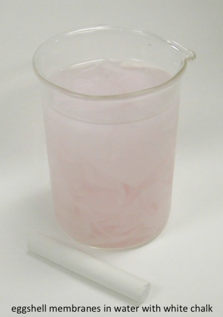
Picture credits: the egg anatomy diagram is from Wikipedia; the confocal image of the fluorescein maleimide-labeled eggshell membranes was obtained by Sonali Raje, Colin Thorpe and Kirk Czymmek; the still life – eggshell membranes with reclining chalk – was by CT.
